Respiratory system of a cat
[
The main function of the respiratory system is to effectively supply the blood with oxygen. Breathing also provides thermoregulation by removing excess water. The normal body temperature of a cat is higher than that of people, somewhere around 38-39°C, and in kittens it can reach 40°C. The expansion of the chest by the action of the pectoral muscles and the arching of the diaphragm create negative pressure in the chest, due to which the lungs inflate and draw air through the nose, and during physical activity through the mouth. The breathing rate in cats ranges from 20 to 30 breaths per minute; in young cats this figure is higher and can reach 40 breaths. The respiratory organs of cats include: nose, nasopharynx, bronchi, trachea and lungs.
The air that a cat inhales passes through the olfactory apparatus of the nose, surrounded by the frontal sinuses, where it is warmed, moistened and filtered. Through the pharynx, which belongs to both the respiratory and digestive tracts, air passes into the larynx and reaches the lungs through the trachea.
The larynx consists of a cartilaginous tube that prevents food from entering the trachea and is involved in sound production due to the vibration of the vocal cords located in it. The cause of the ear-pleasing cat purr is not fully understood. Presumably these sounds arise with the help of the so-called pocket-shaped folds, also located in the larynx.
The trachea is a straight cartilaginous tube that is constantly kept open by C-shaped cartilage. The “open” part of the cartilage is attached to the esophagus, which allows food bolus to pass through it. When a cat eats, the trachea is covered by the epiglottis, and the nasal cavity is covered by the soft palate. Inside the lungs, the trachea is divided into two bronchi: the main and lobar, which in turn, like branches on a tree, are divided into many bronchioles, ending in air sacs or alveoli. The blood circulating around the alveoli is saturated with oxygen.
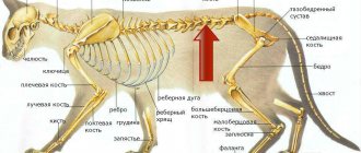
The lungs of cats have the shape of a truncated cone with an apex in the region of the first ribs and a concave base that corresponds to the dome of the diaphragm, and are divided into 2 parts - the left and right lung. Each of them, in turn, is divided into 3 lobes: the upper cranial, middle and the largest lower caudal. The left lung has an additional lobe, making it slightly larger than the right. The volume of the right lung is on average 8 cubic cm, and the left - 11. In their structure, the lungs are similar to a bunch of grapes, where the alveoli are the berries.
Cardimopathy
Among all disorders of the heart, cardiomyopathy is most often diagnosed in cats of various age groups and breeds. Pathology caused by structural abnormalities in the muscular structures of a hollow organ inevitably leads to disruption and dysfunction of the pumping system. In severe cases, congestive heart failure develops, which will lead to anemia and oxygen starvation.
Due to the accumulation of fluid in the pulmonary circulation, respiratory distress syndrome develops. Leads to the formation of blood clots that clog blood vessels. In cats, edema, paralysis, paresis of the hind limbs, acute anemia, and fainting are noted. If treatment is not started in time, the disease is fatal in 100% of cases.
Most cardiopathies in animals are of primary origin. The secondary form of the disease develops rarely and can be caused by surges in blood pressure, anemia, and hyperthyroidism.
In veterinary medicine, there are four types of primary cardiomyopathies in cats:
- Hypertrophic. Characterized by thickening of the myocardium, increased pressure in the cavities of the heart. Leads to the development of acute heart failure.
- Restrictive. With this pathology, there is a loss of elasticity of the myocardium, which leads to degeneration of organ tissue. The systolic and diastolic function of the heart muscle is impaired.
- Dilatational. Characterized by thinning and stretching of the heart walls. The heart enlarges and its contractile function is impaired.
- Arrhythmogenic. A rare genetically determined disease characterized by the replacement of normal tissues with fibrofatty tissue. The pathological process mainly involves the right ventricle.
Each disease has its own specific symptoms and manifestations. Some diseases have a genetic, hereditary origin. Thus, hypertrophic cardiomyopathy is most often noted in Sphynx cats, Norwegian Forest cats, British cats, Maine Coons, Siamese, Abyssinian cats, as well as Redgall and Scottish Fold breeds.
Symptoms
Behavior changes in cats with cardiomyopathy. The condition may be stable until the pathology reaches a critical stage. The disease is manifested by weakness, drowsiness, breathing problems, and unstable temperature. Pulse and blood pressure changes.
Sharp changes in emotional state are noted. Bouts of calm are replaced by increased excitability. Cats meow pitifully, demanding the attention of their owners, hiding in secluded places.
Treatment
Treatment of cardiomyopathies in cats depends on the form and severity of the disease and is most effective at an early stage of development. Therapy is selected individually, in each specific case.
Used in treatment:
- beta blockers;
- medications to prevent thromboembolism;
- blood thinners;
- diuretics;
- calcium channel blocking drugs;
- vitamins;
- homeopathy.
If cardiomyopathy of any form and etiology is detected, the animals, despite their breed qualities, are removed from breeding.
Cat's circulatory system
[
Cats do not have any special differences from the circulatory systems of most mammals. A cat's pulse can be measured by pressing on the femoral artery, located on the inside of the thigh. In normal condition, a cat's pulse is 100-150 beats per minute. And in kittens, the pulse, as well as temperature and breathing rate, is much higher than in adult animals.
As the heart pushes blood through the arteries, their elastic walls actively contract and relax. This is called the pulse. Veins have thinner walls than arteries, so they are more susceptible to damage. There is no pulse in the veins, but the blood moves through them strictly in one direction - towards the heart - due to the valves located in the veins.
Different parts of the body need different amounts of blood. For example, the brain accounts for only a small part of the body's weight, but it requires 15-20% of the total blood contained in the body. Muscles at rest consume about 40% of the blood, and during physical activity (chasing prey, fleeing from a rival or enemy), up to 90% of all blood can circulate in them, that is, blood can be directed to the muscles even from the brain.
Arteries carry bright red blood from the heart throughout the body, enriched with oxygen in the lungs and nutrients in the digestive system. The veins carry darker blood laden with carbon dioxide to the lungs, liver and kidneys.
The exceptions are the pulmonary artery and pulmonary vein. The pulmonary arteries and their capillaries carry oxygenated blood to the pulmonary alveoli, where oxygen is absorbed from the air the cat inhales. The pulmonary veins return fresh blood to the heart, which pumps it through arteries throughout the body. Oxygen enters the cells in exchange for carbon dioxide, and veins carry waste blood to the heart to be pumped back into the lungs for oxygenation.
Veterinarian house call
Treatment and prevention
Self-medication if a cat has a heart disease, regardless of the degree of complexity, can lead to serious consequences. Therapy should be prescribed by the attending veterinarian, having the diagnostic results in hand. The choice of methods depends on the form, stage, complexity of cardiovascular pathology, as well as on the age, physiological state of the animals, and the root cause.
Important! If the pathology is detected at the initial stage of development, the animal is registered with the veterinary clinic. In the future, the veterinarian-cardiologist monitors the treatment and condition of the sick patient.
Treatment of most heart diseases in the early stages of development involves drug therapy. Cardiac glycosides, anticoagulants, drugs that normalize heartbeat, blood pressure, diuretics, restoratives, and symptomatic drugs in injections or tablets are used. Animals are prescribed a special diet, medicinal feed, enzyme agents, vitamins, mineral supplements, and immunomodulators.
The main goal of cardiac therapy is to normalize the functioning of the heart, stop destructive processes in the organ, improve the condition of blood vessels, prevent the formation of blood clots, and correct blood pressure. For each individual pathology, appropriate treatment and certain medications are prescribed.
Sick pets require optimal living conditions and a complete fortified diet. Food should contain proteins, taurine, vitamin A, B3, B6, B12, calcium, phosphorus, magnesium, iron, essential amino acids. It is very important to protect cats from stress, which not only disrupts the functioning of the heart, but also weakens the body.
In severe cases, to eliminate anatomical defects, if drug treatment does not produce results, surgical treatment is used, after which maintenance therapy is prescribed. If it is not possible to completely restore organ function, cats are prescribed lifelong treatment.
Cat's heart
[
The cat's heart, like the human heart, is essentially a twin pump for pumping blood. The body of an average Abyssinian cat (weighing about 3.2 kg) contains just over 200 ml of blood. With each beat, 3 ml of blood passes through the heart. In cats, the heart has a structure similar to that of the hearts of other mammals, but is slightly smaller in size relative to the size of the body.
Through the circulatory system, blood enters the right side of the heart, which pumps it out through the pulmonary artery to the lungs for oxygenation. Already oxygenated blood disappears from the lungs to the left side of the heart. Next, the heart pumps it into the aorta, from where it spreads throughout the cat’s body.
Each part of the heart has an upper chamber - the atrium, and a lower chamber - the ventricle, which is the main pump that forces blood. The tricuspid (or atrioventricular) valve prevents blood from returning to the right atrium from the right ventricle when it contracts. The mitral valve performs a similar function on the left side of the heart. The ventricular muscles, connected to the valves by tendons, prevent them from being pushed towards the atria when the ventricles contract.
Diagnosis of cardiac pathologies
It is impossible to independently determine cardiac pathology in a pet without experience and special equipment. Heart disease, even in its early stages, may not manifest itself for several years. This is why it is very important to take your pet at least once a year for a checkup.
The diagnosis is made taking into account the combination of a number of studies, which include:
- ECHO (echocardiography).
- ECG (electrocardiography). The electrical activity of the heart is measured.
- MRI, CT.
- Tonometry.
- Physical research.
- X-ray. This technique allows you to determine the size and shape of the heart.
- Laboratory tests (serological studies).
In addition to the basic techniques, the doctor collects medical history data and takes into account the specifics of clinical manifestations.
Mechanism of disease development
There are several types of pathologies in the heart area. The first type is valve disease, characterized by a decrease in the amount of blood entering the body due to blood leaking through the valve itself. The second type of pathology is myocardial diseases, characterized by a general weakening or thickening of the heart muscle, which results in a decrease in the efficiency of blood pumping.
There is no single common cause for the development of heart pathologies. But problems with diet are more likely to lead to cardiovascular disease. In addition, the mechanism for the development of heart disease is based on the general physical condition of the animal. Thus, cats that are overweight are more often susceptible to pathological conditions of the myocardium.
The age of the pet plays an important role, because the older the pet, the higher the likelihood of developing cardiomyopathies. Some cat breeds, such as American Shorthairs, Maine Coons and Persians, have a genetic predisposition to developing heart disease.
Alarming symptoms
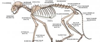
Normally, the anatomy of a cat's heart is not very different from a human's, except for the size of the organ, and it consists of 4 chambers. The pet's internal organ is located above the ulnar tubercle. Owners are not always able to recognize heart pathologies on time, since many diseases do not show any clinical picture for a long time.
With myocardial disease, the animal is forced to stick out its tongue while breathing.
Most heart pathologies are characterized by the following symptoms:
- decreased motor activity;
- lethargic and apathetic state;
- constant desire to sleep;
- rapid breathing, in which the cat takes more than 40 breaths per minute;
- shortness of breath not provoked by physical activity;
- protruding tongue when breathing;
- problems with appetite;
- pale and blue visible mucous membranes;
- tachycardia, in which the heart beats rapidly.
Causes
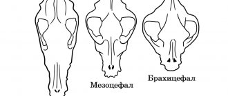
Epilepsy attacks in cats do not have a breed or gender predisposition. But statistically, more cases are registered in exotic animals, where a modification of the skull is observed, and cats suffer from this pathology more often than females. There is no explanation for this - just statistics. The disease can be transmitted genetically, but not necessarily directly from one generation to another, maybe through a generation or two.
The most important causes of epilepsy are disturbances in the conduction activity of nerve impulses in the brain. They are acquired during life, or cats are born with them. Various additional factors act as provocations of attacks. These factors are cerebral (structural pathologies of the brain) and extracerebral (toxic effects on the brain without primary changes in its structures).
- Structural changes in the brain: vascular pathologies, hemorrhages, inflammatory processes with edema, trauma, neoplasms, etc.
- External factors: poisons and toxins, hypoxia (oxygen starvation), jumps or drops in blood enzyme levels, etc.