General structure
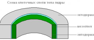
The hydra's body looks like an oblong sac, the walls of which consist of two layers of cells - ectoderm and endoderm.
Between them lies a thin gelatinous non-cellular layer - mesoglea, which serves as a support.
The ectoderm forms the covering of the animal’s body and consists of several types of cells: epithelial-muscular, intermediate and stinging.
The most numerous of them are epithelial-muscular.
Ectoderm
epithelial muscle cell
Due to the muscle fibers lying at the base of each cell, the hydra's body can contract, lengthen and bend.
Between the epithelial-muscle cells there are groups of small, round cells with large nuclei and a small amount of cytoplasm, called intermediate cells.
When the hydra's body is damaged, they begin to grow and divide rapidly. They can transform into other types of cells in the hydra body, except for epithelial-muscular ones.
The ectoderm contains stinging cells that serve for attack and defense. They are mainly located on the tentacles of the hydra. Each stinging cell contains an oval capsule in which the stinging filament is coiled.
Structure of a stinging cell with a coiled stinging thread
If prey or an enemy touches a sensitive hair located outside the stinging cell, in response to irritation the stinging thread is ejected and pierces the body of the victim.
Structure of a stinging cell with discarded stinging thread
Through the thread channel, a substance that can paralyze the victim enters the victim’s body.
There are several types of stinging cells. The threads of some pierce the skin of animals and introduce poison into their bodies. The threads of others are wrapped around the prey. The threads of the third are very sticky and stick to the victim. Usually the hydra “shoots” several stinging cells. After the shot, the stinging cell dies. New stinging cells are formed from intermediate ones.
How do freshwater hydras feed?
Hydra is a predator; its food is ciliates, small crustaceans - daphnia, cyclops and others; sometimes it comes across larger prey in the form of a mosquito larva or a small worm. Hydras can even cause harm to fish ponds by eating fish fry that hatch from the eggs.
Hydra hunting is easy to observe in an aquarium. Having spread its tentacles wide so that they form a trapping net, the hydra hangs with its tentacles down. If you watch a sitting hydra for a long time, you can see that its body is slowly swaying all the time, describing a circle with its front end. A cyclops swimming past touches the tentacles and begins to fight to free itself, but soon, struck by stinging cells, it calms down.
The paralyzed prey is pulled up to the mouth by the tentacle and devoured. During a successful hunt, the small predator swells with swallowed crustaceans, whose dark eyes shine through the walls of the body. Hydra can swallow prey larger than itself. At the same time, the predator’s mouth opens wide, and the walls of the body stretch. Sometimes part of the out-of-place prey sticks out of the hydra's mouth.
The structure of the inner layer of cells
Endoderm lines the entire intestinal cavity from the inside. It consists of digestive muscle and glandular cells.
Endoderm
Digestive system
There are more digestive muscle cells than others. Their muscle fibers are capable of contraction. When they shorten, the hydra's body becomes thinner. Complex movements (movement by “tumbling”) occur due to contractions of muscle fibers of ectoderm and endoderm cells.
Each of the digestive-muscle cells of the endoderm has 1-3 flagella. Vibrating flagella create a current of water, which drives food particles towards the cells. Digestive muscle cells of the endoderm are capable of forming pseudopods, capturing and digesting small food particles in the digestive vacuoles.
The structure of the digestive muscle cell
Glandular cells in the endoderm secrete digestive juice into the intestinal cavity, which liquefies and partially digests food.
The structure of the glandular cell
Prey is captured by the tentacles using stinging cells, the venom of which quickly paralyzes small victims. By coordinated movements of the tentacles, the prey is brought to the mouth, and then, with the help of body contractions, the hydra is “put on” the victim. Digestion begins in the intestinal cavity (cavitary digestion) and ends inside the digestive vacuoles of the epithelial-muscle cells of the endoderm (intracellular digestion). Nutrients are distributed throughout the hydra's body.
When the digestive cavity contains remains of the prey that cannot be digested, and waste products of cellular metabolism, it contracts and empties.
Breath
Hydra breathes oxygen dissolved in water. She has no respiratory organs, and she absorbs oxygen over the entire surface of her body.
Circulatory system
Absent.
Selection
The release of carbon dioxide and other unnecessary substances formed during life processes is carried out from the cells of the outer layer directly into the water, and from the cells of the inner layer into the intestinal cavity, then out.
Nervous system
Below the skin-muscle cells are star-shaped cells. These are nerve cells (1). They connect with each other and form a nerve network (2).
Nervous system and irritability of the hydra
If you touch the hydra (2), then excitation (electrical impulses) occurs in the nerve cells, which instantly spreads throughout the entire nervous network (3) and causes contraction of the skin-muscle cells and the entire body of the hydra shortens (4). The response of the hydra body to such irritation is an unconditioned reflex.
Intestinal cavity
Let's look at the internal structure of the hydra in more detail. The hydra's body looks like an oblong sac. Its walls consist of two layers of cells, between which there is an intercellular substance (mesoglea). Thus, there is an intestinal (gastric) cavity inside the body. Food enters it through the mouth opening. It is interesting that the hydra, which is not currently eating, has practically no mouth. The ectoderm cells close and grow together in the same way as on the rest of the body surface. Therefore, every time before eating, the hydra has to break through its mouth again.
The structure of the freshwater hydra allows it to change its place of residence. On the sole of the animal there is a narrow opening - the aboral pore. Through it, liquid and a small bubble of gas can be released from the intestinal cavity. With the help of this mechanism, the hydra is able to detach from the substrate and float to the surface of the water. In this simple way, with the help of currents, it spreads throughout the reservoir.
Sex cells
With the approach of cold weather in the fall, germ cells are formed from intermediate cells in the ectoderm of the hydra.
There are two types of germ cells: eggs, or female germ cells, and sperm, or male germ cells.
The eggs are located closer to the base of the hydra, sperm develop in tubercles located closer to the mouth.
The egg cell of a hydra is similar to an amoeba. It is equipped with pseudopods and grows rapidly, absorbing neighboring intermediate cells.
The structure of the hydra egg cell
The structure of the hydra sperm
Spermatozoa resemble flagellated protozoa in appearance. They leave the hydra's body and swim using a long flagellum.
Radiation symmetry
Let's take a closer look at the external structure of the hydra. The table will help you remember the parts of the body and their purpose.
| Part of the body | Purpose |
| Intestinal cavity | Digestion, movement |
| Mouth | Penetration of food |
| Tentacles | Food capture, defense, movement |
| Foot | Attachment to substrate |
| Aboral time | Detaching from the surface |
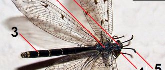
The body of the hydra, like many other animals leading an attached lifestyle, is characterized by radial symmetry. What it is? If you imagine a hydra and draw an imaginary axis along its body, then the animal’s tentacles will diverge from the axis in all directions, like the rays of the sun.
The structure of the hydra's body is dictated by its lifestyle. It attaches itself to an underwater object with its sole, hangs down and begins to sway, exploring the surrounding space with the help of tentacles. The animal is hunting. Since the hydra lies in wait for prey, which can appear from any direction, the symmetrical radial arrangement of the tentacles is optimal.
Fertilization. Reproduction
The sperm swims up to the hydra with the egg cell and penetrates inside it, and the nuclei of both sex cells merge. After this, the pseudopods are retracted, the cell is rounded, a thick shell is released on its surface - an egg is formed. When the hydra dies and is destroyed, the egg remains alive and falls to the bottom. With the onset of warm weather, the living cell located inside the protective shell begins to divide, the resulting cells are arranged in two layers. From them a small hydra develops, which comes out through a break in the egg shell. Thus, the multicellular animal hydra at the beginning of its life consists of only one cell - an egg. This suggests that the ancestors of Hydra were single-celled animals.
Asexual reproduction of hydra
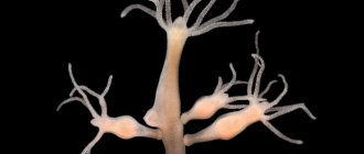
Under favorable conditions, hydra reproduces asexually. A bud forms on the animal’s body (usually in the lower third of the body), it grows, then tentacles form and a mouth breaks through. The young hydra buds from the mother's body (in this case, the mother and daughter polyps are attached with tentacles to the substrate and pull in different directions) and leads an independent lifestyle. In autumn, hydra begins to reproduce sexually. On the body, in the ectoderm, gonads are formed - sex glands, and in them, germ cells develop from intermediate cells. When hydra gonads form, a medusoid nodule is formed. This suggests that the hydra gonads are highly simplified sporifers, the last stage in the series of transformation of the lost medusoid generation into an organ. Most species of hydra are dioecious; hermaphroditism is less common. Hydra eggs grow rapidly by phagocytosing surrounding cells. Mature eggs reach a diameter of 0.5-1 mm. Fertilization occurs in the body of the hydra: through a special hole in the gonad, the sperm penetrates the egg and merges with it. The zygote undergoes complete uniform fragmentation, as a result of which a coeloblastula is formed. Then, as a result of mixed delamination (a combination of immigration and delamination), gastrulation occurs. A dense protective shell (embryotheca) with spine-like outgrowths is formed around the embryo. At the gastrula stage, the embryos enter suspended animation. Adult hydras die, and the embryos sink to the bottom and overwinter. In the spring, development continues, in the parenchyma of the endoderm, an intestinal cavity is formed by divergence of cells, then the rudiments of tentacles are formed, and a young hydra emerges from under the shell. Thus, unlike most marine hydroids, hydra does not have free-swimming larvae and its development is direct.
How do hydroids differ from other coelenterates?
Hydroids are the most simply structured coelenterates with a clearly defined two-layer structure.
- This is the only class whose colonies can include both polyps and jellyfish.
- Only among representatives of this class of cnidarians there are freshwater species.
- Hydroid polyps never have septa in the gastric cavity, and their nervous system does not form ganglia or sensory organs.
- In hydroid jellyfish, unlike scyphoid jellyfish, the radial canals are unbranched.
- Hydroid nematocysts lie only in the epidermis (ectoderm), but the diversity of their stinging cells is great.
- There are no cells in the mesoglea.
- Gonads from germ cells are formed either in the gastrodermis or in the epidermis.
- In general, they are characterized by alternating generations of jellyfish (sexual generation) and polyp (asexual generation). But in many species, one of these generations (usually the jellyfish) is suppressed or completely absent. In the latter case, the remaining generation (usually a polyp) becomes sexual.
- The skeleton (not everyone has it) is secreted outward by the ectoderm in the form of a cuticle, sometimes impregnated with calcium.
- In hydroid jellyfish, along the edge of the umbrella there is a special membrane - a sail (vellum) .
- On the tentacles of jellyfish there is one type of sensory organs: either only statocysts (most often), or eyes, eyespots, or complex visual organs with lenses.
 Statocysts are organs of balance; they are closed vesicles lined with sensitive epithelium and filled with fluid. Inside each bubble (jellyfish have them there are pieces of calcium carbonate that act as statoliths.
Hydroid Far East
Regeneration
Hydra has a very high ability to regenerate. When cut crosswise into several parts, each part restores the “head” and “leg”, maintaining the original polarity - the mouth and tentacles develop on the side that was closer to the oral end of the body, and the stalk and sole develop on the aboral side of the fragment. The whole organism can be restored from individual small pieces of the body (less than 1/100 of the volume), from pieces of tentacles, and also from a suspension of cells. Moreover, the regeneration process itself is not accompanied by increased cell division and is a typical example of morphallaxis.
Reflexes
The structure of the hydra is such that it is able to sense changes in temperature, the chemical composition of water, as well as touch and other stimuli. The nerve cells of an animal are capable of being excited. For example, if you touch it with the tip of a needle, the signal from the nerve cells that sensed the touch will be transmitted to the rest, and from the nerve cells to the epithelial-muscular cells. The skin-muscle cells will react and contract, the hydra will shrink into a ball.
This reaction is a clear example of a reflex. This is a complex phenomenon consisting of successive stages - perception of the stimulus, transfer of excitation and response. The structure of the hydra is very simple, therefore the reflexes are monotonous.
Movement
In a calm state, the tentacles extend several centimeters. The animal slowly moves them from side to side, lying in wait for prey. If necessary, the hydra can move slowly.
"Walking" mode of transportation
"Walking" method of movement of the hydra
Having curved its body (1) and attached its tentacles to the surface of an object (substrate), the hydra pulls the sole (2) to the front end of the body. Then the walking movement of the hydra is repeated (3,4).
Description and lifestyle
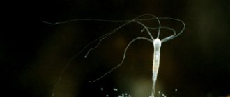
A common inhabitant of water bodies, the freshwater polyp called hydra is a coelenterate. It is a gelatinous translucent tube up to 1 cm long. At one end, on which a peculiar sole is located, it is attached to aquatic plants. On the other side of the body there is a corolla with many (6 to 12) tentacles. They are capable of stretching up to several centimeters in length and are used to search for prey, which the hydra paralyzes with a stinging injection, pulls with tentacles to the oral cavity and swallows.
The basis of nutrition consists of daphnia, mosquito larvae, fish fry, and cyclops. Depending on the color of the food eaten, the color of the hydra’s translucent body also changes.
Thanks to the contraction and relaxation of integumentary muscle cells, this organism can narrow and thicken, stretch to the sides and move slowly. Simply put, the most similar thing to a moving and independent stomach is the freshwater hydra. Its reproduction, despite this, occurs at a fairly high rate and in different ways.