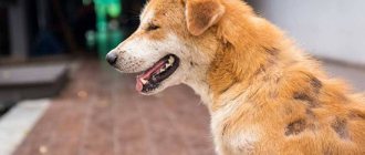
Eye diseases in pets pose a particular danger to their health. This is due to the fact that four-legged pets receive most of the information about the world around them through vision. Beginning dog breeders cannot always distinguish conjunctivitis from glaucoma, let alone inflammation of the third eyelid in dogs. Many of them do not know at all that the pet has this very third eyelid. The article will discuss in detail the functions of the nictitating membrane, the causes of its pathology, as well as treatment methods.
What is the third eyelid?
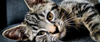
The popular name “third eyelid”, scientifically in veterinary medicine is called the crescent-shaped fold of the conjunctiva; it is located in the corner of the eyeball.
The absence of pigmentation on the free edge of the third eyelid is not a pathology, but only a feature of some animals.
The semilunar fold protects the eye from various injuries. If you press lightly on your dog's eye, it will immediately appear and instantly cover the surface of the cornea. The second feature is that in the thickness of the third eyelid there is an additional gland, which ensures the production of about 30% of tears. Also, when the third eyelid moves, tears are evenly distributed over the surface of the cornea, which helps remove or wash away foreign particles and bacteria.
Diagnostics
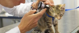
To collect a complete medical history, the doctor interviews the dog's owner and also conducts a physical and ophthalmological examination, which includes testing the pupils and measuring eye pressure. Additionally, the following diagnostic measures may be prescribed:
- Examination of the third eyelid using tweezers under local anesthesia.
- Examination by a neurologist.
- Taking a general blood test.
- X-ray examination for the presence of a bony orbit.
- Ultrasound of the eye and surrounding tissues inside the orbit.
- Computed tomography of the brain, eyeball, bone orbit.
- Magnetic resonance imaging (MRI).
Diseases of the third century
With the insanely fast pace of life, a person sometimes does not notice some of the changes that occur to his pet and lead to various diseases. This does not happen with diseases of the third eyelid; probably the very first place the owner will look is the eyes. Even minor changes will be noticeable immediately. Therefore, we will consider the most common diseases in dogs.
Volvulus
Occurs in young dogs up to one year old. This is due to the fact that in an unformed body, cartilage sometimes grows faster than the rest of the body, so the edge of the cartilage pushes out the third eyelid, and an incorrect anatomical position of the cartilage is observed towards the bend. There is a disruption in the normal functioning of all tissues of the eyelid, which leads to swelling, redness and hyperplasia (increase in the number of cells).
The most common problems are Newfoundland, Central Asian Shepherd, and Great Dane.
Treatment is carried out using surgery, excision of the cartilage occurs. Healthy, undamaged cartilage is not subject to excision.
Inflammation
As a rule, inflammation is caused by the action of pathogenic and conditionally pathogenic microorganisms, but in addition to this, there are a number of factors that cause this disease:
- damage, loss of integrity, eye injuries;
- getting small objects and dust into the eyes;
- irritation to smoke, various chemicals;
- with prolonged and uncontrolled administration of potent drugs;
- after a number of diseases, for example carnivore plague.
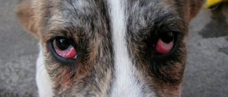
The owner can easily determine the signs of the inflammatory process. The dog has a slight prolapse of the eyelid, it is very hyperemic and has swelling, the size varies from a millimeter to a centimeter. The animal feels pain when touched near the eyelid, often lies down and tries to hide from bright light.
Treatment: use anti-inflammatory drugs, antiseptics. If the disease manifests itself as a result of infection, treatment is carried out with antibiotics and antiviral drugs.
Adenoma (true and false)
This is a common ophthalmological disease, which is characterized by a benign tumor neoplasm.
It is often confused with hyperplasia due to similar clinical signs - the organ enlarges and extends beyond its normal anatomical location, this is a false adenoma.
A true adenoma is confirmed by biopsy; if it confirms the presence of a tumor, then surgical treatment is used. The operation is determined by the doctor. If the tumor is small, does not interfere with the animal and does not limit the function of the eye, then surgical intervention is not required.
Hunting and decorative breeds are more susceptible to adenoma.
Hyperplasia
Hyperplasia is a pathological growth of tissue of the third eyelid. The clinical picture is very similar to an adenoma, only in this case the tissue proliferation is not caused by tumor processes. The nictitating membrane is enlarged, the dog’s third eyelid is red and falls out a little. The causes of hyperplasia have not been fully established. In order to treat hyperplasia, you need to know its etiology; treatment is generally conservative, but in some cases surgical intervention is resorted to.
Loss (prolapse)
A characteristic sign is the third eyelid closing some part of the eye (half, a third of the eye). The cause of prolapse is often a weakening of the thin ligament that holds the third eyelid. Hair loss is not always an independent disease; sometimes it is a consequence of serious illnesses. This pathology can develop in the following cases:
- foreign body;
- eye injury;
- entropion of the eyelid;
- inflammatory processes or malignant/benign neoplasms of the bones of the upper jaw, nose, eye orbits.
Loss may occur in stressful situations (especially in small breeds), hidden infections, or parasitic diseases. As well as dogs suffering from ectropion of the eyelid (eversion of the eyelids).
The breeds most prone to prolapse are: pug, Chinese Crested and Mexican, American and English Cocker Spaniel, Yorkshire Terrier, English Bulldog, Cane Corso, Chihuahua.
Lacrimal gland prolapse
This is a pathological phenomenon in which the lacrimal gland is displaced outward, pinched by the edge of the third eyelid. Dogs most susceptible to the disease are: Pekingese, French/English bulldog, Shar Pei, Cane Corso, Cocker Spaniel, Pug, Toy Terrier, Chihuahua.
This is due to congenital hyperplasia of the lacrimal gland of the third eyelid and weakness of the ligament that holds it. In normal condition, the lacrimal gland is covered by the third eyelid, but with allergic reactions, chronic conjunctivitis, or breed predisposition, it enlarges and falls out. Next, it is pinched, which causes redness and swelling of the tissue.
Without medical intervention, purulent or mucous discharge from the eyes, dryness, swelling, or squinting may occur.
It is treated surgically, the gland is set and anti-inflammatory and antibacterial eye drops are prescribed.
Third century gap
Occurs only under mechanical stress; all breeds of dogs are susceptible. Clinically, this may appear as a small tear that will not disturb the pet. Or a rupture with a piece of drooping eyelid that will need to be promptly removed. Minor tears can heal on their own; the rest requires surgical intervention by an ophthalmologist.
Follicular conjunctivitis
Accompanied by inflammation of the lymphatic follicles, which are located on the inner surface of the third eyelid and on the conjunctiva.
The cause is chronic inflammation, irritation from dust, smoke, chemicals, and allergies. The size of the conjunctivitis follicle ranges from 0.5 to 3 mm. Signs of advanced stages of follicular conjunctivitis:
- squinting of eyes;
- lacrimation, mucus-like discharge;
- redness and swelling of the conjunctiva;
- noticeable follicles on the conjunctiva.
Treatment: If the disease is not caused by an infection, then eye drops with corticosteroids can be prescribed.
If the dog’s body does not respond to drug treatment, then surgical intervention is used: they are cauterized with an electrocoagulator or a chemical agent, and the follicles of the third eyelid are scraped out.
In dogs, follicular conjunctivitis of the third eyelid must be differentiated from plasmoma.
Contacting a veterinarian and treatment
Diagnosis may require blood tests, ultrasound, x-rays, MRIs and biopsies. Based on the data obtained, medications are selected for the four-legged patient or surgery is prescribed.
Drug therapy
Treatment of inflammation of the third eyelid in dogs with the help of drugs only relieves swelling. It is used for small swellings, absence of itching and preservation of the size of the formation.
To eliminate the primary disease, antibiotics, antivirals or glucocorticosteroids are used. The site of inflammation is treated with antiseptics and anti-inflammatory agents to prevent suppuration.
Operation
Surgical treatment is provided for adenoma, volvulus, eversion, prolapse, lymphatic conjunctivitis and severe rupture. Depending on the situation, the eyelid is sutured or adjusted.
Removal of the membrane and gland is resorted to only in the most severe cases. Due to the reduction in the amount of tear fluid after surgery, animals often develop keratoconjunctivitis sicca. To prevent it, it is necessary to regularly moisturize the mucous membrane with an “artificial tear” preparation.
First aid
It is important to provide first aid to your pet before contacting a specialist, which will help reduce pain and slow down the development of the inflammatory process.
It is recommended to use the following drugs:
Dexamethasone is an eye drop that has an anti-inflammatory effect and does not cause an allergic reaction. Instill 2 drops in the morning and evening into the third eyelid area. Do not use in the presence of purulent formations.
Tsiprovet is an antibiotic that will eliminate the inflammatory process of an infectious nature. Instill 1 drop four times a day.
Dexamethasone - eye drops to relieve inflammation.
Important! Do not self-medicate. At the first symptoms of diseases of the third eyelid, you should contact a veterinarian to avoid serious pathologies.
Disease prevention
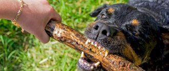
Prevention of inflammation of the third eyelid will help prevent the disease or significantly reduce the risks. Often the disease occurs as a result of mechanical damage to the eyes, so it is important to monitor your pet during games and walks. This is especially true for moving breeds - hunting and guard dogs.
When buying a dog from a breeder, you need to carefully study the characteristics of the breed and possible diseases. It is worth considering heredity. If the puppy's parents had similar diseases, the risks increase.
If preventive measures do not help, at the first symptoms you should consult a doctor and not select medications on your own. Advanced disease leads to blindness.
Read Signs and treatment of ataxia in dogs: 3 types of disease
Briefly about the main thing
- The main functions of the third eyelid are to protect the cornea and remove foreign particles.
- In most cases they are treated surgically.
- For effective treatment, the type of disease must be determined.
- Adenoma is often confused with hyperplasia, but it is extremely rare.
- Treatment at home can lead to vision loss.
- Removing the third eyelid will not cause severe discomfort to the animal.
- Removing the lacrimal gland can cause serious consequences, including loss of vision.
Removal
There is no drug treatment program for problems with the third eyelid. The only way out is surgery to remove the third eyelid.
Purpose of surgery:
- do not disturb the anatomical structure of the third eyelid,
- preserve the mobility of the eyelid as much as possible;
- exclude the development of the disease in order to preserve the animal’s vision.
What does the eyelid look like after surgery?
The specialist will inform you about the need to remove cartilage or part of the lacrimal gland only as a last resort, if the consequence of maintaining the tumor may be loss of vision.
The operation is performed under general or local anesthesia, is not complex, and does not require a long recovery period.
How to feed after surgery
After the operation, to prevent the dog from injuring himself with his paw, he needs to wear a protective collar . During the first 7-10 days after surgery, you should prevent the development of bacteria by using antibacterial medications for the eyes.
You should not trust the operation to a novice veterinarian. Resection errors can cause functional disorders of the eyeball.
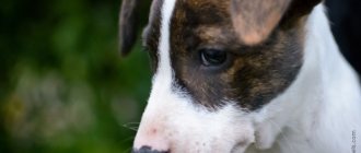
Main symptoms
It is quite easy to identify diseases because the symptoms are characteristic. An oblong or round formation is observed near the medial canthus. The shade varies from pink to bright red.
The enlarged gland is then damaged when blinking, injuring the eye cornea. Uncomfortable sensations appear, which is why the pet begins to scratch the problem area. This leads to infection and damage. Follicular hypertrophy also develops, purulent-mucous discharge and lacrimation appear.
Initially, the pathology is observed in one eye, but after one to three months the second eye may be affected. If the necessary medical intervention does not occur, there is a risk of developing dry eye syndrome and pigmentary keratitis.