To understand why tumors appear in cats after sterilization, you need to understand what operations to “disable” the reproductive function in females are. In modern veterinary clinics such procedures are carried out in one of the following ways:
- laparoscopic;
- classical.
Laparoscopic .
Through small (up to 1 cm) incisions on the abdomen, a laparoscope (tube with a video camera) and a manipulator are inserted into the abdominal cavity. Carbon dioxide is supplied to raise the abdominal wall so that the surgeon can see all the organs. Directly in the abdominal cavity, the ovaries and uterus are removed and removed through incisions, bleeding vessels and tissues are cauterized with a laser or electric current. The cat requires virtually no post-operative care. If the incisions are smaller than 0.5 cm, they are not sutured, but sealed with medical glue. Classic method . The incision is made along the linea alba of the cat's abdomen. The aponeurosis (wide tendon plate) of the abdominal wall is dissected, the muscles are not affected. The surgeon removes the ovaries and uterus, ligates the vessels and cuts off the organs. The peritoneum is sutured with special threads, which dissolve on their own after a certain time. Such seams do not require any special care; during this period the main thing is to ensure that the cat cannot reach them.
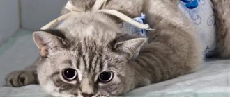
During the rehabilitation period after sterilization, lymphatic fluid may accumulate in the cavity formed under the external suture, forming swelling visible to the naked eye. This symptom usually appears 2–5 days after surgery. The “tumor” does not cause discomfort to the cat, does not require treatment and resolves on its own.
Post-operative swelling is normal and should not cause any alarm to the pet owner. If the appearance of swelling near surgical sutures is observed after laparoscopy or is accompanied by suspicious symptoms, consultation with a veterinarian will be required.
Alarming symptoms
What signs of deterioration in health in an animal that has recently undergone sterilization should alert a caring owner? First of all, the cat owner should suspect something is wrong if, more than 2 days after the operation, his pet still shows signs of abdominal pain:
- screams a lot and loudly;
- refuses food;
- if he lies, then only on his side or on his back.
Another alarming sign is a temperature that does not subside for a long time. Normally, surgery provokes an immune response in the cat’s body, as a result of which the thermometer consistently shows a value greater than 39°C for 3 days after the operation, which is completely normal.
However, if the tailed patient’s temperature does not decrease either on the fourth or fifth day after sterilization, this is already a clear sign that an inflammatory process is underway in the body, and the lump that has formed on the pet’s belly is its direct consequence.
After sterilization, the following symptoms may appear:
- allergic reactions (rash, swelling of the mucous membranes);
- lethargy, drowsiness;
- arrhythmia;
- loose stools or constipation;
- urinary incontinence;
- enlargement of the mammary glands;
- wheezing breathing;
- bleeding of sutures.
Sometimes more than one of the above symptoms may occur. As mentioned earlier, signs of the inflammatory process are increased body temperature and redness of the skin. If you do not contact a veterinarian in time, complications can lead to the death of the animal.
Important! Post-surgery lumps are often confused with a mammary tumor in a cat. There is a difference between them. In most cases, the lump goes away on its own. The tumor only grows over time, and similar tumors form next to it.
Reasons for appearance
There are many reasons for the appearance of lumps on a cat’s belly after sterilization. Let's look at the most common of them:
- Postoperative swelling. This is a safe and harmless reason for the formation of a lump after surgery. It occurs for physiological reasons due to the healing of tissue damaged as a result of the incision. It is worth noting that despite the possible impressive size (up to 5 centimeters in diameter), the formation itself does not cause the animal any discomfort. The swelling subsides on its own within 3–5 days after surgery.
- Hernia. The formation of a postoperative hernia is a more dangerous phenomenon that can lead to significant health problems for the animal. The cause of its occurrence is the divergence of the suture of the abdominal wall. This is due to the fact that in the process of making the incision, the veterinarian has to cut through several layers of tissue - the skin, the subcutaneous fat layer, the muscles of the abdominal wall and, in fact, the peritoneum itself. After the operation is completed, tissue stitching occurs in the reverse order. Sometimes it happens that the suture on the peritoneum is made incorrectly, as a result of which it diverges and a small gap is formed into which the cat’s internal organs fall out. They protrude, which on the outside looks like a lump or lump. Postoperative hernia is a complex pathology, the treatment of which involves repeated surgery.
- Ligature fistulas. They are formed as a result of inflammation and suppuration of non-absorbable threads that were used to make a surgical suture. At first they may look like small bumps on your cat's belly. The bumps very quickly develop into pustules, which are foci of infection. The disease requires repeated surgical intervention in rare cases; usually treatment is conservative, and it is aimed at eliminating the infectious process.
- Accumulation of lymph. If the lymphatic vessel is poorly sutured, the lymph may leak and accumulate, forming a lump. Over time, it resolves on its own or is removed with the help of drainage.
My animals had just an accumulation of lymph. The accumulated fluid was pumped out of the dog with a syringe. In cats, the cones resolved on their own within 2 weeks.
When to see a doctor
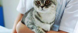
During the recovery period after sterilization, it is recommended to show the cat to a veterinarian. How painlessly and quickly she recovers largely depends on this.
Of course, lumps, which are postoperative edema, are quite physiological. In this case, contacting a veterinarian is not required. However, if the lump has a large diameter, a rash, redness appears around it, and ichor or pus is released, then this indicates the seriousness of the pathology and requires prompt seeking veterinary help.
At what age are cats spayed?
The ideal age for sterilization in cats is a relative concept. The operation can be performed at almost any age; from 6 months to 10 years it is tolerated quite easily. Older individuals may have contraindications or complications. Today, many veterinarians recommend carrying out the procedure up to a year or after the first heat. At a young age, the animal tolerates all manipulations easily and quickly adapts to the new state. He has not yet formed stable habits, which can be stronger than hormones. For example, an adult that is accustomed to marking its territory will likely continue to do so after spaying or neutering. Therefore, it is wise to act on warning and form the pet’s character and habits from a young age. It is believed that by 8 months a cat reaches sexual maturity and can bear and bear offspring up to 4 times a year. But, firstly, this is individual, for some individuals this moment comes earlier, for others later, and secondly, there are breeds in which the offspring grow for a long time and mature only by one and a half to two years. It is too early to sterilize such cats at 8 months, and it is better to select the timing of the operation together with a veterinarian. In general, the bulk of procedures occur between the ages of one and three years.
What to do if a lump forms on your stomach
As mentioned above, if the swelling does not subside for several days, increases in size, festeres, or the pet feels unwell, be sure to contact a veterinarian to avoid serious complications. The veterinarian, having established the cause, will prescribe symptomatic treatment aimed at normalizing the general condition of the operated animal.
When a postoperative hernia forms, if there is no pinching, the problem is solved by repeated sutures or reduction of the hernial protrusion. If the process is started, surgery may be required.
If the tumor is malignant or benign, the veterinarian, after tests, decides on the choice of treatment. The animal is prescribed maintenance therapy or surgery.
In any case, the choice of treatment methods for the appearance of a lump on the abdomen, above or below the suture, depends on the root cause, the general condition of the cat, and diagnostic results.
Important! Do not self-medicate so as not to harm your mustachioed pet. All manipulations and therapy should be prescribed by a veterinarian.
Treatment and prevention
If, after sterilization, the cat has a lump under the stitch, it lasts for several days, pus has formed, the cat has become irritable or apathetic, you should immediately consult a doctor.
Let us note the various treatment options:
- After the operation, a hernia appeared. In this case, the veterinarian evaluates the animal’s condition and may prescribe a second operation or correct the hernia.
- Pus has formed. In a clinical setting, the pus is removed and medications are prescribed to stabilize it. In extreme cases, a repeat operation will be required. Veterinarians will often prescribe ointments or a special blanket to support the pet's abdomen. This way the stitches will not come apart and the pet will not be able to lick the sore spot. This procedure will protect against re-infection.
- Serious inflammation. In most cases, it is better for the animal to remain in the clinic for a while under the supervision of doctors. Sooner or later the pet will return home.
Let us note the basic recommendations that doctors prescribe:
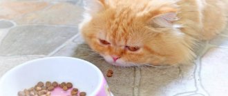
- Special diet. Much depends on the breed and nature of the disease. Veterinarians often recommend special collars to limit the cat's food intake. You can use specialized dry food for animals that have undergone surgery. Portions should be small, food (if it is natural food) easily digestible. It is better to grind the food and keep hard pieces to a minimum.
- The diet when a seal appears on the seam and after its removal is extremely important. Any problems with the gastrointestinal tract - constipation or excessive gases can greatly affect the dehiscence of the suture. If the cat starts vomiting, this is a sign of recovery from anesthesia. It is worth limiting your pet's food intake and providing plenty of fluids.
- Increased attention. Don't bother your cat unnecessarily. Protect her from drafts and contact with other animals. improve your sleeping area. For example, put new litter in a cat's nest. If necessary, provide favorite toys and clean drinking water. It is better to place the bowl next to the sleeping place.
- Required activity. The animal can sleep a lot, but periodically make it move. A supervised walk around the apartment would be an excellent choice. Avoid active games with your cat, and do not let her play with her favorite toys for a long time.
The main thing is not to delay treatment if a lump appears under the stitch.
IMPORTANT! It is important to know that this may not just be a hernia, but a serious cancer. Under no circumstances should you self-medicate.
If a seal appears on the seam, consultation with a veterinarian is required.
What should you do if your cat has a lump on her stomach after sterilization? How is this dangerous for the health of your beloved pet and is swelling a postoperative complication? What triggers the formation of a lump in the scar area?
Therefore, if your cat has a round, dense swelling in the tissue incision area, and you are worried about this, we recommend reading this review, from which you will learn what can cause a “bump” on the abdomen (under the suture), what it means, and what to do in such a situation.
Preventing the appearance of a lump on the stomach
In order for the postoperative period to pass calmly and without the development of complications such as the appearance of a lump on the abdomen, you should carefully follow all the veterinarian’s prescriptions.
Make sure that for the first two days the cat does not jump from a height or show excessive activity. Coming off of anesthesia, reflexes are dulled, the body is weakened, and behavior is unpredictable. Therefore, do not leave your pet uncontrolled.
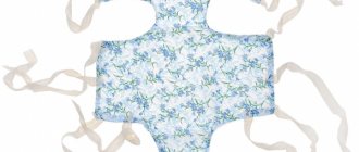
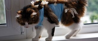
The seam should be regularly treated with disinfectants until it is completely healed. To prevent the cat from licking it and tearing out the threads, a blanket is put on it. Some animals manage to get out of this “clothing”; for such cats it makes sense to use a protective collar.
As soon as your cat shows interest in food, feed her small portions. Food should be nutritious and easily digestible. You can give special foods intended for feeding during the postoperative period or formulations for sterilized, neutered animals.
Proper prevention and systematic proper care will help the cat return to normal faster after sterilization. If there are no complications and you follow all the veterinarian’s recommendations, the rehabilitation period will take no more than five weeks.
After sterilization, a cat has a lump under the seam: what to do
In any case, complications are reported to the veterinary surgeon. If postoperative swelling has developed, the swollen surface is treated with an antiseptic spray that forms a protective film. The condition of the swelling is monitored, and re-treatment is carried out if the need arises.
If a significant amount of lymph is released, the veterinarian installs drainage or prescribes a wound-healing powder. The abscesses are opened, the wounds are treated with antimicrobial external agents.
If a hernia has formed, a repeat operation is performed.
Cesarean section for cats
Cesarean section for cats
Caesarean section (sectio caesarea) is an emergency obstetric operation to pull the fetus through an incision in the abdominal wall and uterus, performed when it is impossible to remove it through the natural birth canal, as well as in case of fetal asphyxia. The purpose of this operation is to relieve the fetus and mother while maintaining her productivity. The term Sectio caesarea has been used in medicine since ancient times. This method of assisting in the birth of a child was first legalized by the Roman Caesar Numa Pompilius (713-673 BC), allowing the fetus to be removed through an incision in the abdomen in the event of the mother’s death during childbirth, and the operation received the name “caesarean section” because With her help, Julius Caesar was born.
Operations used to remove dead fetuses, tumors and other indicators do not qualify as a caesarean section.
Caesarean sections are performed much more frequently in cats than in other types of pets. This is explained by the simplicity and speed of the operation, the high recovery rate and the difficulty of performing routine obstetric care in these animals due to the narrowness of the birth canal and the long length of the uterine horns.
Indications: cesarean section is performed in cases of cervical fusion, uterine torsion, periostitis and abnormalities in the mother's pelvic cavity, abnormal positions of the fetuses, overdeveloped fetuses, deformities and abnormalities of fetal development.
According to other authors, indications for surgery in cats are frequent indications for cesarean section: discrepancy between the volume of the birth canal and the volume of the fetus, bending of the fetal head and neck, and absence of labor. Uterine torsion in carnivores is relatively rare and is segmental, so it occurs only during laparotomy and is not an indication for cesarean section.
Prognosis: the success of the operation depends on the timing of its implementation
The sooner a cesarean section is performed from the onset of pathological labor, the more favorable the prognosis. A dark green discharge from the cat's vulva indicates that labor began several hours ago; they indicate separation of the placenta and possible fetal death. The chances of recovery are reduced with fetal emphysema. In this case, it is important to establish the viability of the uterine tissue; if they have necrobiotic changes, a hysterectomy is performed.
A fetus trapped in the vagina rarely remains alive for more than 8 hours from the onset of the second stage of labor due to placental abruption. On the contrary, fetuses in the uterus can remain alive for up to 36, and sometimes up to 48 hours. Typically, a caesarean section can be performed safely within 12 to 24 hours. After the start of the gestation stage. If the operation is performed later than 24 hours. From the beginning of the fetal stage of labor, the probable death of the fetus with infection of the uterus. In this case, a hysterectomy is performed with removal of the uterus at the level of the vagina and cervix to reduce the possibility of infection of the abdominal cavity.
After a cesarean section, the female is fertilized, but there is a high probability that the next birth will also be pathological. The prognosis, of course, depends on the operative technique. A.P. Students, V.S. Shipilov, L.M. Subbotina recommend performing the operation as follows.
Online access. Depending on whether the fetuses are alive, different surgical approaches are chosen: medial, pramedial or oblique paralumbar
For live fetuses in cats, it is better to use a median incision.
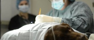
Anesthesia. The operation is performed on a lying anesthetized animal.
When prescribing anesthesia, the age and condition of the woman in labor are always taken into account. Before performing an operation, it is advisable to inject an animal that is in a serious condition intravenously with a 5% glucose solution, a 10% calcium gluconate solution, cordiamine, and then, immediately before administering anesthesia, a 0.5% atropine solution is injected.
Preparation of the surgical field: depilation is carried out on the abdominal wall, the skin is lubricated twice with an iodine solution and the field is covered with sterile L-shaped napkins or a piece of linen with an oval notch 15–20 cm long. The animal is placed in a dorsal position. It is more convenient to make the incision along the white line between the last two nipples.
Technique for performing caesarean section in cats
The median surgical approach begins 2 cm from the navel and continues along the linea alba for a length of 7–10 cm in cats, approximately to the level of the last nipples of the mammary gland. In this case, the skin, subcutaneous fatty tissue, and aponeuroses of the oblique and transverse abdominal muscles are dissected. If, when removing the uterus, the length of the incision is insufficient, then it is continued in the cranial direction, bypassing the umbilicus.
The paralumbar surgical approach passes in the area of the right ventricle from top to bottom and forward parallel to the course of the muscle fibers of the internal oblique muscle. It starts in front of the knee fold (2-3 fingers), runs parallel to the last rib. The transverse abdominal muscle is separated with the handle of a scalpel following the course of its fibers, then the transverse abdominal fascia and peritoneum are cut.
The omentum is pushed to the side and, carefully, inserting the index fingers under the uterus in the area where the fetus is located and grasping the accessible anterior part of the uterine horn, slowly remove it out. The uterine horn is covered with napkins and dissected near the body of the uterus by 7–10 cm. To prevent severe bleeding, you should not dissect the uterus from the side and especially near the lesser curvature. For the same reasons, incisions in the placental area are avoided. The latter is easy due to the larger volume and thickness of the uterine wall. In this case, uterine discharge should not enter the abdominal cavity. If it is difficult to remove all the fruits through one cut, cut another corner. In cases where the uterus contains many fetuses, two incisions are made approximately in the middle of each horn.
The fetuses are removed from the uterus in membranes with one hand, bringing them to the incision and squeezing them out through the wall of the uterus with the second hand
Together with the fetus, after uniform tension along the umbilical cord, the placenta also departs. The umbilical cord is torn and the fetus is transferred to an assistant, who immediately ruptures the membranes and frees the fetal airways from mucus, dries the body with a napkin and places the fetus in a warm place. For fetuses delivered via caesarean section, heat is very necessary.
Other authors indicate that the wound of the horns should be located near the body of the uterus: this makes it possible to extract the fruits from both horns through one incision
A number of authors note that in order not to prematurely disrupt the integrity of the membranes, the horn is dissected after first opening it between tweezers in the same way as is done when cutting the peritoneum. The edges of the horn wound are grabbed with tweezers and the fruits are removed through the incision.
Some authors note that it is better to obtain the fruits together with the shells; if the shells rupture, then one of the most important elements of the operation after extracting the fruits is the rapid removal of water with gauze swabs or suction. The fetuses in the membranes are immediately transferred to a special assistant. It quickly breaks the fruit shells, wipes the front of the head, mouth and frees the oral and nasal cavities from mucus; thoroughly dry the skin with napkins and tie the umbilical cord.
Other authors point out that if artificial respiration is necessary, it is performed immediately, bending the newborn’s body. A number of authors point out that heat has a particularly beneficial effect on newborns: they are placed in a thermostat or wrapped in gauze napkins and cotton wool, and covered with heating pads.
After the uterus is freed from the fetus, bacteriostatic agents are introduced into its cavity.
When bleeding from the mucous membrane, it is useful to lightly compress the uterus with gauze compresses. The edges of the wound are sutured in layers.
The uterine cavity is washed with a warm solution of furatsilin, the remains of the placenta are removed, especially in the case of late cesarean section for fetal emphysema; gloves are also washed. After this, the uterine cavity is dried with tampons and 50,000-100,000 units of penicillin are injected into it.
A Schmieden suture is applied to the uterus with catgut No. 4-5 and, after clearing of blood clots and washed with furatsilin, a Lambert suture is applied with the same catgut. After repositioning and straightening the uterus and intestines, the cat is injected into the abdominal cavity with 50,000 units of penicillin or another antibiotic dissolved in a 0.5% solution of novocaine. To prevent adhesions, oil solutions of antibiotics (mastisan, mastidine, etc.) or mild fibrinolytics are used. The abdominal cavity is closed with a continuous catgut suture or a separate loop of similar stitches. A continuous catgut suture is applied to the peritoneum with muscle aponeuroses or directly to the muscles (depending on the laparotomy access), and a knotted silk suture is applied to the skin.
Often the wound is left open, smearing its edges with a 5% alcohol solution of iodine. Sometimes the wound is sprinkled with tricillin powder on top, the ends of the threads are pulled apart in directions opposite to the wound, a gauze cloth is placed on the wound and tied with skin suture threads. It is also practiced to apply an adhesive (collodion or BF-6) bandage.