Quite often, veterinary specialists are faced with diagnosing leukoma (eye sore) in patients. The pathological process is direct evidence of the onset of the inflammatory process in the transparent protective layer of the eyeball - the cornea.
Neglect of the disease and untimely assistance can lead to serious problems in the cat such as blindness or complete loss of the eyeball.
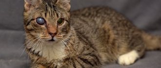
Leukoma or cataract is a clouding of the cornea of the eye with the formation of a scar. The development of leukoma is provoked by such negative factors as thermal and chemical burns, allergic reactions, and mechanical injuries. White connective tissue begins to grow after damage, impairs the cat's vision and can lead to complete blindness.
The cat's eyesore, like other animals, is divided into several types. It depends on the location of the white film. There are peripheral leukoma, which covers the edge of the eye (peripheral vision is impaired), as well as central (develops in the center) and total (affecting the entire visual surface).
The inflammatory process that develops with leukoma causes the scar to rise above the cornea.
What it is
Leukoma in cats is a pathological condition characterized by the appearance of a scar on the cornea, the formation of which can be caused by various ophthalmological diseases.
What does leukoma indicate?
As the disease progresses, changes occur in the organ of vision. The animal begins to behave differently: hide from the light, look for secluded places.
The cat's tear production increases.
Consequences of ignoring the problem
If dangerous changes are not detected in a timely manner, the cataract will degenerate into adipose tissue. This condition can lead to complete atrophy of the eye. In addition, the disease causes a lot of inconvenience to the cat.
The resulting film provokes a decrease in visual acuity, especially when localized on the pupil itself.
White film growth
The third eyelid in a healthy cat is visible only when blinking, in a state of sleep.
Importance for a cat
The formation protects the cornea from dust, debris, plant thorns, etc., evenly distributes the secretion of the lacrimal gland, and protects the surface of the eye from drying out.
Reasons for sinking
An increase in film size can be associated with both external and internal factors.
There are several reasons for the development of a pathological condition:
- bacterial, viral, fungal infections;
- failure of internal organs;
- exhaustion, dehydration;
hormonal imbalance;- entry of foreign bodies;
- oncology;
- weakening of the ligaments and muscles of the eye;
- helminthiasis;
- injuries;
- allergic conjunctivitis;
- atrophy of the eyeball.
British and Persian cats are more susceptible to the development of vision pathologies due to the structural features of the skull. Regardless of other reasons, genetic predisposition is a fundamental factor in the occurrence of the disease.
Signs of adenoma
This is a benign formation. It is characterized by the appearance of a small tumor that prevents the eyes from closing completely.
Symptoms:
increased body temperature;- purulent discharge from the eyes;
- photophobia;
- neoplasms in the corners of the eyes;
- loss of appetite;
- irritability.
The animal also continuously washes itself, squints, and blinks.
Associated symptoms
If the cause of the adenoma is systemic diseases, the following signs of ill health are observed: vomiting, diarrhea, weakness, decreased activity, weight loss, deterioration in coat quality.
Types of pathology
An eyesore is classified according to several criteria.
By location type
Highlight:
- peripheral;
- total;
- central.
In the first case, the pupil is not affected; the spot is mainly localized on the side of the eyeball, has a white tint and black inclusions.
With the total type, the cataract completely covers the entire eye.
In the central form, the center of the visual organ is affected, and complete occlusion of the pupil is often noted.
According to the pathological process
Leukoma can be congenital or acquired . The first type of disease is rarely diagnosed and occurs against the background of negative changes in the cat’s body that occurred during the period of intrauterine development.
The second form is more common and can be caused by many factors.
Eye diseases in humans: list, symptoms
The reason for this is many factors. For example, the rapid development of computer technology and the deterioration of the environmental situation every year. Next, we will consider the most common diseases, and also highlight their characteristic symptoms.
Pathology of the optic nerve
Glaucoma
- a chronic disease. Due to increased pressure inside the eyes, optic nerve dysfunction occurs. As a result, vision decreases, which may disappear in the future. The disease progresses very quickly, so the patient risks completely losing his vision if he delays going to the doctor. Signs: impaired lateral vision, black spots, “hazy” images, inability to distinguish objects in the dark, colored rings appear in bright light.
Ischemic optic neuropathy
– circulatory disorders in the intraocular or intraorbital region. Symptoms: decreased visual acuity, appearance of “blind” spots in some areas. Reducing viewing angle.
Ischemic neuropathy
Neuritis
- infection. An inflammatory process in the optic nerve is characteristic. Signs: loss of sensitivity in the area around the eye, pain, weakening of the muscles associated with the optic nerve.
Nerve atrophy
– a disease characterized by dysfunction of arousal conduction. Color perception and viewing angle are impaired. Vision decreases and a person can become completely blind.
Nerve atrophy
Pathology of the eye orbit, eyelids, lacrimal canals
Blepharitis
- inflammation that occurs along the edges of the eyelids. Symptoms: swelling of the tissue, accompanied by burning and redness. The patient feels as if a speck has gotten into his or her eye. There is itching and characteristic discharge. Bright light is difficult to perceive, tearing, pain. Dry eyes and peeling of the eyelid margins may occur. After sleep, purulent scabs form on the eyelashes.
Blepharitis
Cryptophthalmos
- a common disease in which the edges of the eyelids fuse together. This causes the palpebral fissure to narrow or even disappear.
Lagophthalmos
– a pathology characterized by a violation of the closure of the upper and lower eyelids. As a result, some areas remain open all the time, including during bedtime.
Turn of the century
– the place where eyelashes grow is turned towards the eye socket. This creates severe discomfort due to rubbing and irritation of the eyeball. Small ulcers may form on the cornea.
Turn of the century
Coloboma of the century
- disturbance in the structure of the eyelids. Usually occurs along with other morphological defects. For example, cleft palate or cleft lip.
Swelling of the eyelid
– localized accumulation of excess fluid in the tissues around the eyelid. Symptoms: local redness of the skin, discomfort. Eye pain worsens when touched.
Swelling of the eyelid
Blepharospasm
- looks like a convulsive contraction of the facial muscles, as if the person is quickly squinting his eyes. Not controlled by the will of the patient.
Ptosis
– drooping of the upper eyelid. Pathology is classified into several subtypes. In some cases, the eyelid droops so much that it completely covers the eyeball.
Ptosis
Barley
– an infectious disease of an inflammatory nature that occurs with pus discharge. Signs: swelling of the edges of the eyelids, redness and peeling. Pressing is accompanied by severe pain. Discomfort (feeling of a foreign object in the eye) and lacrimation are common. The acute form is characterized by signs of intoxication - loss of strength, fever, headache.
Barley
Trichiasis
– improper eyelash growth. The danger is that pathogens can easily enter the eyes. This provokes inflammation, conjunctivitis and other problems.
Dacryocystitis
– an infection of the tear duct that causes inflammation. There are several types of pathology: acute, chronic, acquired, congenital. Symptoms: painful sensations, the lacrimal sac is red and swollen, suppuration of the canals and constant tearing.
Dacryocystitis
Pathology of the tear-producing system
Dacryodenitis
- damage to the lacrimal glands. It occurs due to chronic pathologies, or due to infection entering the body. If there is a disruption in the functioning of the circulatory system, the disease can take a chronic form. Symptoms: the upper eyelid becomes red and swollen. In some cases, the apple of the eye protrudes. If dacryodenitis is not treated, the inflammation spreads, ulcers form, a high temperature rises, and general malaise appears.
Dacryoadenitis
Lacrimal gland cancer
– develops as a result of abnormal activity of gland cells. Tumors can be either benign or malignant. The second group includes, for example, sarcoma. Signs: pain in the eyes and head. Associated with an increase in formation that puts pressure on the nervous tissue. In some cases, the pressure is so strong that it causes delocalization of the eyeball, making it difficult for them to move. Additional symptoms include swelling and loss of vision.
Pathology of the connective membrane of the eye
Xerophthalmia
– an eye disease during which tears are produced less than normal. There are several reasons for this: chronic inflammatory processes, various injuries, tumors, long-term use of medications. Elderly people are at risk.
Conjunctivitis
- inflammation that occurs in the conjunctival mucosa. It can be allergic, infectious and fungal. All of these varieties are contagious. Infection occurs both through physical contact and through everyday objects.
Tumors of the conjunctiva
– appearing in the coal on the inner side of the mucosa (pterygium) and forming in the area of the connection with the cornea (pinguecula).
Lens pathology
Cataract
– gradual clouding of the eye lens. The disease develops very quickly. It can affect one eye or both. In this case, either the entire lens or one part is damaged. The main category of patients is elderly people. It is this disease that can reduce vision in a very short time, even to the point of blindness. In young people, cataracts are possible due to injuries or somatic diseases. Symptoms: rapid loss of vision (this forces you to change lenses very often), inability to distinguish objects in the dark (“night blindness”), impaired color perception, eyes get tired quickly, and in rare cases, double vision.
Cataract
Lens abnormalities
– cataracts, bifaf, spherophakia, lens luxation, coloboma developing from birth.
Retinal pathology
Retinitis (retinal pigmentary dystrophy)
– a disease manifested by the occurrence of inflammation in various parts of the retina. The causes include injury to the organs of vision and prolonged exposure to sunlight. Symptoms: the normal field of vision narrows, visibility decreases, the image doubles, insufficient visibility at dusk, characteristic colored spots appear before the eyes.
Retinal detachment
– a pathology in which destruction of the retina is observed. Its inner layers begin to peel away from nearby epithelial tissues and blood vessels. In most cases it is treated surgically. Lack of treatment results in vision loss. Signs: “fog” before the eyes, distortion of the geometric shape of objects, sometimes flashes of light and bright sparks flash through.
Retinal detachment
Retinal angiopathy
– destruction of the structure of the choroid in the eyes. This disease is caused by physical trauma, high intraocular pressure, disturbances in the functioning of the central nervous system, diseases of the circulatory system (arterial hypertension), poisoning, and pathological defects in the morphology of blood vessels. Symptoms: noticeable decline in vision, blurred vision, foreign flickers, image distortion. In the most severe cases, vision loss occurs.
Retinal dystrophy
– an extremely dangerous disease that can have a wide variety of causes. The tissue of the retina of the eye dies or decreases. This can happen if qualified assistance from specialists is not provided in a timely manner.
Corneal pathology
Keratitis
– an inflammatory process that affects the cornea of the eye. As a result, clouding of the cornea and the occurrence of infiltrates. The cause may be an infection: viral, bacterial. Injuries can also trigger the development of the disease. Symptoms: lacrimation, redness of the mucous membrane of the eye, atypical sensitivity to bright light, the cornea loses its normal properties - shine, smoothness. If treatment is neglected, the infection spreads to other areas of the visual system.
Keratitis
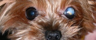
Belmo
– formation of scar tissue on the cornea of the eye, its persistent clouding. The cause is prolonged inflammatory processes in the body or injury.
Belmo
Corneal astigmatism (keratoconus)
– degeneration of the cornea, which occurs due to increased pressure inside the eye. This leads to a change in the shape of the cornea of the eye. Symptoms: light fringe around the light bulbs, immediate decrease in vision in one of the eyes, myopia.
Keratoconus
Change in eye refraction
Myopia (myopia)
– a refractive error in the eye, in which a person has difficulty seeing distant objects. In case of myopia, the image is fixed in front of the retina. Signs: poor discrimination of distant objects, discomfort, rapid eye fatigue, pressing pain in the temples or forehead.
Myopia
Farsightedness (hypermetropia)
– a refractive error in which the image is read behind the retina, is the opposite of myopia. In this case, the patient has difficulty seeing both near and distant objects. Symptoms: very often there is blurriness before the eyes, sometimes the patient exhibits strabismus.
Farsightedness
Astigmatism
– the disease is characterized by the inability to focus light rays on the retina. Usually appears in people with physiological disorders of the visual organs: cornea, lens. Symptoms: blurred and unclear image, a person gets tired quickly, often complains of a headache; in order to see something, one has to strain the eye muscles.
Astigmatism
Other eye diseases
Nystagmus
– uncontrollable oscillatory movements of the eyeballs.
Lazy eye syndrome or amblyopia
– a pathology in which the eye, due to damage to its muscles, stops working and making movements.
Anisocoria
– difference in pupil size. Basically, it appears with all kinds of eye injuries. Involves acute sensitivity to light and decreased vision. Sometimes this pathology indicates a disruption in the functioning of one of the parts of the brain - the cerebellum.
Anisocoria
Episcleritis
- inflammation that forms in the episcleral tissue. First, redness appears near the cornea, then this area swells. Signs: feeling of discomfort, eyes hurt from bright light. There are discharges from the connective membrane. In most cases, episcleritis goes away on its own.
Episcleritis
Aniridia
– complete absence of the iris of the eye.
Aniridia
Polycoria
– an eye defect when a person has several pupils.
Polycoria
Ophthalmoplegia
– a disease when the nerves of the eye that are responsible for its movement cease to function correctly. This causes paralysis and the inability to rotate the eyeballs. Symptoms: eyes are turned to the nose, do not change this position.
Exophthalmos
– pathological exit of the eyeball beyond the orbit of the eye, occurs due to swelling of its tissue. In addition to the main symptoms, redness of the eyelids and pain when touching the inflamed area are noted.
Diplopia
– a disorder of the visual system, consisting of constant double vision of visible objects.
Causes of cataracts
Various factors can contribute to the formation of a cloudy film and stain. The problem in kittens is identified in rare cases when the cat suffered from infectious diseases at the time of birth.
Among the main reasons that provoke a thorn are the following:
- dysfunction of the endocrine system;
- eye injury;
- failure to comply with hygiene rules;
- infections;
- unbalanced diet.
Diseases in which ocular leukoma develops
Veterinarians identify some pathologies that can lead to clouding and the appearance of a white spot on the visual organ.
Trachoma
This is a disease of infectious nature. In most cases, eye damage is accompanied by the formation of a cataract.
Inflammation on the cornea
One of the most common provoking factors of the disease.
If veterinary care is not provided in a timely manner, the risk of pupillary clouding increases several times. The superficial form is characterized by a weak degree of severity, and the thorn is almost invisible.
Rehabilitation after treatment
In most cases, treatment for leukoma is quite long and takes 2-3 months. After drug treatment, it is advisable to carry out preventive methods in the form of taking antiviral drugs, vaccination, and deworming. A proper, balanced diet is essential - preferably using high-quality, veterinarian-approved food. You also need to monitor the animal during walks and other active activities to avoid eye injuries.
During the postoperative period, the animal is prescribed antibacterial therapy, some restriction of physical activity, and mandatory medical supervision over the recovery process.
Symptoms of pathology
It is difficult to treat a thorn. It is necessary to pay attention to changes in a timely manner and show the cat to a specialist.
The pathological process is accompanied by the following symptoms:
increased lacrimation;- fear of light;
- swelling;
- redness of the cornea;
- discharge of purulent contents;
- retinal atrophy;
- development of strabismus.
Depending on the type of lesion, the clouding may have a light yellow or red-gray uneven tint. In the ulcerative form of the disease, the thorn protrudes outward.
In most cases, the animal’s behavior changes noticeably: the cat becomes more aggressive and irritable, is unsure of its orientation in space, bumping into objects that appear in its path, washes itself frequently,
Preventive measures
To protect the tailed creature and minimize the occurrence of problems with the visual apparatus, you should do the following:
- plan a cat’s diet - it must be balanced and consist of quality products;
- do not forget about additional vitamins in the cat’s menu;
- periodically rinse your cat's eyes with Furacilin (1 tablet per 100 g of water);
- use potassium permanganate for preventive purposes;
- regularly examine the eyes of the mustachioed creature and monitor changes in its behavior;
- do not forget to free your pet from roommates - worms, fleas, etc.
- Take your pet to the veterinary clinic for examinations at least once a year.
If you notice slight redness of the mucous membrane and tearing, use Tricilin powder on the affected areas.
Diagnostics: techniques and methods
During the initial examination, the veterinarian evaluates the clinical picture and conducts an external examination of the pet, especially the eye area.
The specialist also collects information about when the spot was first noticed, the quality of food, and studies the animal’s history.
Based on the data obtained, a diagnostic examination is prescribed, including the following manipulations:
- clinical and biochemical blood test;
- ophthalmoscopy;
- serological tests;
- determining the pressure in the eye (to assess how likely it is to develop glaucoma or cataracts);
- Seidel test (fluorescein test);
cytological examination of damaged tissue taken from the conjunctiva;- biopsy;
- microscopic examination of scraping;
- gonioscopy.
If for some reason it is not possible to conduct a standard diagnostic examination, the doctor may prescribe an ultrasound examination, which will help identify the true causes of clouding of the eyes.
If concomitant diseases of viral and infectious origin are suspected, additional diagnostic methods may be prescribed.
Basic principles of therapy
Treatment of cataract requires strict adherence to all the veterinarian’s instructions.
Medicines
Eye drops are used for 14 hours.
This could be Floxal, Tetracycline. In the morning and evening, it is necessary to apply a product with antibacterial properties to the lower eyelid. It is advisable to do the procedure at the same time.
When the provoking factor of the lesion is injury, the eye is first treated with an antiseptic (Miramistin), followed by the use of drops, for example, Levomycetin.
To relieve severe pain, the doctor advises injecting a special solution into the eyeball. For increased eye pressure, Brinzolamide and Pilocarpine are prescribed.
To obtain the desired effect, all medications must be used in combination.
Surgical intervention
If drug therapy does not help, surgery is performed.
During keratoplasty, the cornea is completely replaced. Tarsorrhaphy involves suturing the edges of the eyelid, which will protect the cornea and speed up recovery.
ethnoscience
Treatment of leukoma at home is possible only after a full examination and in combination with medications.
A honey solution is effective. To prepare it, you need to mix honey with water in a small amount and drop 3 drops into your eyes until complete recovery.
Blepharitis
Inflammation of the eyelids, which is quite common in furry cats, is divided into three types:
- Scaly blepharitis (also called simple);
- Ulcerative blepharitis;
- Meibomian blepharitis.
Symptoms differ for each type, but there are common ones that are always noticeable. Namely:
- Itching;
- Edema;
- Redness;
- Tearing.
With scaly blepharitis, you can notice a clear thickening of the eyelids, after which gray scales appear there. Over time, the eyelashes will begin to fall out, and pus will be released in place of the scales.
If treatment is not started, scaly blepharitis can quickly turn into ulcerative blepharitis. With it, crusts of pus dry out, fall off, and in their place sores open. The inflammation gets worse. Scarring may cause the skin to tighten.
Meibomian blepharitis is similar to a stye. The eyelid swells and turns red. Pus begins to ooze out profusely.
Preventing eye problems in cats
To prevent the development of the disease, it is important to adhere to a number of simple rules:
provide the animal with healthy, nutritious nutrition;- regularly examine your eyes;
- protect your pet from contact with other animals;
- do not let outside unattended;
- carry out vaccinations in a timely manner.
It is also necessary to regularly visit the veterinarian as a preventative measure, which will allow the disease to be identified at the beginning of its development.
First aid
Self-medication in this case is strictly prohibited. Medicines are used only after consultation with a veterinarian. If you delay in going to the clinic, your pet’s condition will quickly deteriorate.
As soon as the owner notices that his pet has a white film on the organ of vision, and an urgent visit to the veterinarian is impossible, the animal is treated at home.
Wiping the eyelids
Take a clean cotton cloth, moisten it with warm filtered water, and carefully remove all discharge from the eyes from the outer corner to the inner one.
Moisturizing the eyes
If the eye is swollen, carefully spread the eyelids and drip saline solution. If the film falls out, periodically moisten the eye with saline solution and apply a sterile bandage.
Help with bleeding
If bleeding occurs, apply a sterile bandage or gauze folded in several layers to the eye (the material should not stick). When soiled, the bandage is changed, but at least once a day.
Examination by a veterinarian
At the clinic, the doctor conducts a full examination of the pet. An ophthalmoscope is used to examine the condition of the fundus.
If there are no injuries, clinical tests are prescribed.