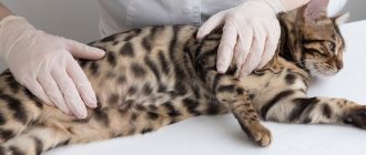
How to examine a cat?
Quite often, the appearance of tears in a cat's eyes indicates the presence of some kind of disease. It is important for the owner to pay attention to whether tears appeared for a limited time and then disappeared, or in the pet’s eyes the discharge is present throughout the day, or, what is much worse, not for several days.
- If the tear discharge is insignificant, appeared in the morning and quickly passed, then there is nothing to worry about, so there is no need to worry;
- If it is clear that the eyes “cry” for a long time, then you need to look for the cause of this phenomenon.
Before examining your pet, you need to clean your hands to prevent additional infection. Next, you need to pull down the upper eyelids on each eye. If the eyes are shiny and not clouded, the pupils are the same size, the cornea is noticeably transparent, and the back surface of both eyelids has a pale pink tint, then there are no problems with the animal’s eyes.
The fact that there are certain problems with the eyes can be judged by redness or swelling of the eyelids, clouding of the cornea.
If the animal’s eyes are difficult to open or there is a thin light film on the inner surface of the eyelid, then it is recommended to go to the veterinarian as soon as possible.
When to contact a veterinarian
Examine the cat carefully. If the tears are clean and transparent, without any admixture of pus or blood, there are no signs of swelling, and the cat plays with pleasure and has an excellent appetite, there is nothing to worry about. Your pet does not require treatment, but only proper care.
A visit to the veterinarian is required if the following signs are detected:
- The cat doesn't eat well.
- Her snot is flowing.
- He often scratches his eyes.
- Cloudy or purulent tears.
- Dull eyes.
- The cat is too excited or, conversely, too lethargic.
- The cat's eye is swollen.
- There are deep scratches and cuts around the eyes.
In specialized veterinary clinics they will always be able to accurately determine the diagnosis and correctly state the cause of watery cat eyes.
Causes and treatment
There are several common reasons why a cat's eyes may start to water.
Some foreign body has entered the eye. It could be a speck, a twig, a fluff, a midge or dirt. If something like this is discovered, you must first rinse the pet’s eyes under running water. Once the foreign object is washed away, you can use one percent tetracycline ointment to apply to the cat's upper eyelid area. It is recommended to take the animal to a veterinarian for consultation.
The pet has a weakened immune system. This happens if the kitten was separated from its mother too early. However, only a veterinarian can reliably determine whether this is the case. He will also develop a regimen for taking the required medications and correct the pet’s nutrition.
The presence of an allergic reaction, which is inherent in cats as well as in humans. The same thing that can act as an allergen can act as a human allergen: chemicals, dust, pollen, etc. In this case, it is imperative to contact a qualified specialist for consultation, since the owner will not be able to independently determine not only the allergy itself, but also the cause of its occurrence.
Worms are a fairly common reason why a cat's eyes may begin to water. To exclude this, it is necessary to regularly treat the animal for parasites.
Pet breed. Some cat breeds have a characteristic skull structure with deformed nasolacrimal ducts and a flattened nose. In this case, only washing the eyes every day helps, as well as special attention to hygiene procedures in such a risky area.
Diseases of cold origin. If your pet coughs and sneezes additionally, absorbs a lot of water, and begins to sleep much more, then it is quite possible to suspect a cold. It is not recommended to treat the animal yourself; you need to contact a specialized specialist.
Cat distemper also has discharge from the eyes as one of its symptoms. In addition, the appearance of pus, high fever, vomiting and diarrhea, and general weakness are noted. It is not allowed to treat a pet with such symptoms on your own.
Treatment of conjunctivitis: how can an ophthalmologist help?
The doctor chooses a treatment strategy depending on the type of conjunctivitis. The disease is treated with medications, rinses and physiotherapy.
Medicines
In case of allergic conjunctivitis, it is necessary to establish the cause and stop contact with the allergen. At the same time, anti-allergy and decongestant drugs, drops to relieve inflammation, vitamins and other general health-improving agents are prescribed.
Bacterial conjunctivitis is treated with antibiotics. They are simultaneously prescribed internally and externally, the course is usually 7-14 days. The infectious type is treated with antiviral, immunostimulating and bactericidal drugs of local (drops, ointments, gels) or general action.
Drug therapy for eye fungi is complex and includes local and general antifungal and immunostrengthening agents. In special cases, IVs are placed.
For injuries or burns, the doctor will prescribe wound-healing, decongestant, antimicrobial agents in the form of drops, sprays, etc. For severe cases of any type of conjunctivitis, a course of injections may be prescribed.
Washing
With conjunctivitis, it is extremely important to maintain good hygiene, so the course of treatment includes frequent eye rinsing. For this purpose, solutions, herbal decoctions with a disinfecting effect, or simply clean water are prescribed.
To carry out the procedure, there are special containers (small oval-shaped glasses) and devices (syringes, bulbs, etc.).
Physiotherapy
Physiotherapy for conjunctivitis involves the administration of drugs using physical methods - current (electrophoresis), laser (laser phoresis), magnet (magnetophoresis), ultrasound (phonophoresis), etc.
Use solutions with anti-allergenic, anti-edematous, antiseptic, wound-healing, softening, anti-inflammatory effects.
Eye diseases accompanied by the production of tears
There are several different eye conditions in pets that cause tear production:
- Infection. If the eye is noticeably swollen and red, and there is discharge of pus, then the help of a veterinarian is definitely needed to prevent the development of feline conjunctivitis.
- Directly eye diseases, in particular glaucoma or cataracts. A clouded cornea in the first case or a whitened lens in the second are the most obvious symptoms of these diseases. Of course, you definitely need to go to a clinic for treatment.
- Diseases caused by fungi or viruses (such as toxoplasmosis or chlamydia) can only be cured by a veterinarian.
- Inflammation of the cornea can be identified by reddened and very watery eyes, on which a soapy film forms.
- Some breeds experience entropion of the eyelid - usually Sphynxes or Persians. The appearance of swollen eyes and suppuration in their eyes is a reason to visit a veterinarian as soon as possible.
- Eye injuries that a cat can get if it fights with its fellow tribesmen, hits itself, or gets caught on some branches. Wounds or other damage to the eyeballs are obvious symptoms of this injury. It is recommended to consult a veterinarian to determine the severity of the damage.
How to distinguish dangerous lacrimation from non-dangerous ones
With pathological
When your cat has watery eyes, problems appear in one or both eyes. The discharge is thin or mucous, brown or slightly whitish.
The nature of the discharge can vary from liquid and transparent to thick, purulent. Typically, pathological lacrimation is accompanied by the animal's anxiety and itching: the cat constantly washes itself and rubs its eyes with its paws.
Body temperature may rise, red eyes, and swelling of the eyelids may appear. Crusts form in the corners of the eyes and hair falls out. When only one eye is watery, the cat squints it and rubs it with its paw. Pathological lacrimation may be accompanied by lethargy of the pet, poor appetite and other signs of illness.
With a sick animal, you should go to the veterinarian to get advice, understand the causes of the disease and take all necessary measures. Timely treatment will help avoid dangerous complications such as infection and blindness.
Tearing in cats can also occur due to
natural
causes. For example, after sleep one eye waters and does not open. This is not dangerous: the eye should be rinsed with boiled water or chamomile decoction.
Long-haired cats often have excessive tearing due to hair getting into the tear duct and the accompanying irritation. Breeders are advised to trim the long hair on the face of a long-haired cat and brush the pet regularly.
Due to the anatomical features of the structure of the skull and the short nasolacrimal ducts in the British, Scots and Persians, lacrimation is a normal variant. These cats cry almost constantly without experiencing any discomfort. Breeders of such cats need to take good care of their pet’s hygiene and periodically treat their eyes and face.
The color of the discharge from the eyes matters
It is important for the pet owner to carefully look at the color of the discharge coming from the eyes.
If they are transparent, similar to ordinary tears, then this may be due to the characteristics of the breed, mechanical irritation, or the occurrence of an allergic reaction.
If yellow or green discharge appears, in which pus is present, we can say that there is an infectious disease.
If a cat's eyes are watery red or brown, then there is a high probability that the cat is infected with worms.
Attention! No self-medication!
The main rule for cat owners is not to self-medicate, no matter how simple the problem may seem. Remember that any wrong action or inaction you take towards your pet can negatively affect its health.
It is always better to contact a competent specialist, for example, to the Murmansk veterinary clinic “Favorite”, where you can always get a full range of consultations, diagnosis and treatment of all cat diseases.
"Favorit" - professional care for your pet!
The Scottish Fold cat's eyes are watery: why?
It was previously mentioned that certain breeds of pets have a non-standard nasolacrimal duct, and among them the British, Scottish and Persian breeds stand out. Due to such features, the lacrimal canal is not able to fully remove secretions, which are the norm for all other breeds. In this case, the excess comes out through the eyes. If this happens after the animal has slept, then there is no reason to worry. This phenomenon is normal from a physiological standpoint.
If your pet constantly produces tears, this is no longer considered normal, so you need to seek the help of a veterinarian to find out the reasons.
Such animals need to wash their eyes once a week or more often, and in some cases up to 2-3 times a day. With a normal level of discharge, it is quite enough to simply wipe them periodically.
If your cat's eyes often water, it is recommended to bring the animal to the veterinarian for a full examination.
Meaning of lacrimation
Tearing has two main functions: hydration and protection. Tears are primarily composed of water and electrolytes, but also contain small amounts of mucus and albumin. Thus, when the tear enters the outer part of the eyeball, it moisturizes the mucous membrane of the eye.
In addition, the production of tears is a natural way of flushing the eyes. With the current of tears, all foreign particles (including dust and microorganisms) are removed from the eye.
Cat protection
A very correct approach is the use of hygienic methods that protect the animal from the need for treatment in the future. It is recommended to periodically wash your pet's eyes with a cotton swab dipped in warm water towards the spout.
If there is dried discharge on the eyes, then you need to carefully soften and remove it, and then rinse the eyes with a clean swab.
You cannot use the same swab to treat both eyes; you need to take a new one each time.
If your pet has watery eyes, you do not need to buy and apply drops or medications yourself without consulting a veterinarian, as this can make the animal sick.
Rules for washing and instilling eye drops
If one of the points of treatment for a cat is eye drops, then it is important to know how to carry it out while causing minimal discomfort to the pet.
For this you need:
- sterile pipette or syringe;
- product (drops, ointment) at room temperature;
- clean wipes or tampons;
- food to encourage and relieve stress in cats.
It is advisable to carry out the burial with an assistant who will help hold the frightened animal. The cat must be well secured. In extreme cases, it is tied with soft cloth.
With a moistened swab, wipe the pet's eye from the outer corner to the inner, removing crusts and drops of secreted liquid. Then the eye is wiped again with a clean napkin.
Now you can start instilling: open the cat’s eyes wide with the thumb and forefinger, after which the product is quickly instilled directly under the eyelid. Excess liquid is blotted with a napkin, then the cat is rewarded for its bravery and released.
Pregnant and small pets require a special approach. Cats expecting the birth of babies are rarely prescribed medications; rinsing is done with chamomile or calendula decoctions, a tea solution, and in extreme cases, tetracycline ointment is applied. Newborn kittens with high tear production are prescribed Bars or Anandin drops.
Preventive procedures
If there is no obvious problem at the moment, you can use the recommendations for prevention. A warm decoction of medicinal plants, which are also intended for humans, drops that do not contain an antibiotic, or green tea are suitable for this.
If your cat's eyes are watery due to an infectious disease confirmed by a veterinarian, then tetracycline ointment can be used.
To treat bacterial infections, “Diamond Eyes” drops or some of their analogues are used.
Viral or bacterial eye infection
A cat's eyes may become watery due to a viral or bacterial infection. If your pet's discharge has been going on for quite some time, and it has become thick, cloudy white or yellow, then most likely there is an infection in the eyes that is rapidly gaining momentum.
This could be conjunctivitis, toxoplasmosis, or herpetic infection. It is necessary to undergo an examination by a veterinarian in Murmansk. Ignoring these alarming symptoms can significantly weaken your cat's vision and even lead to blindness. The specialist will prescribe a series of tests to identify a hidden infection in the body and draw up a treatment plan for your pet.
Bleeding in the eye
Atherosclerosis
Diabetes
Fungus
28656 March 22
IMPORTANT!
The information in this section cannot be used for self-diagnosis and self-treatment.
In case of pain or other exacerbation of the disease, diagnostic tests should be prescribed only by the attending physician. To make a diagnosis and properly prescribe treatment, you should contact your doctor. Hemorrhage in the eye: causes of occurrence, what diseases it occurs with, diagnosis and treatment methods.
Definition
Hemorrhage in the eye is the accumulation of blood under the conjunctiva, in and between the membranes of the eye, in the chambers of the eye or in the vitreous body. Hemorrhage is caused by a violation of the integrity of the blood vessels of the eye.
The eyes continuously perceive, transform and send a huge amount of information to the visual analyzer.
The eyes are sensitive to changes in the body and, although quite protected, are easily damaged by external influences.
The eyeball is located in the cavity of the orbit - a bone frame; the orbit protects the eyeball from above, below, behind and on the sides. The eye in the orbit is located in fatty tissue - a kind of pillow. In front, the eyeball is protected by eyelids and eyelashes.
Eyeball
consists of three shells:
- external, represented by a convex transparent cornea, turning into the sclera - the frame of the eyeball;
- middle - in front is the colored iris, then the ciliary body, and in the back is the choroid itself, represented by a network of small arteries and veins. At the center of the iris is the pupil, which allows light to enter the eye. The ciliary body produces intraocular fluid, which is needed to maintain the shape of the eye and help with metabolism. The lens is attached to the ciliary body with the help of muscles and ligaments. By contracting and relaxing the muscles, the curvature of the lens changes, thus the eye “switches” the distance and focuses the image;
- the inner retina, which contains rods and cones that perceive light and convert it into an electrical impulse, which is then sent to the brain.
Vitreous body
fills the space between the lens and the retina and is a gel-like substance.
The eye has two spaces filled with intraocular fluid: the anterior chamber of the eye (between the cornea and the iris) and the posterior chamber of the eye (between the iris and the vitreous body, which contains the lens).
The cameras communicate with each other through the pupil. Conjunctiva
- This is the mucous membrane, it lines the inside of the eyelids and extends to the visible part of the eye. The conjunctiva is nourished by a network of very small vessels located directly underneath it. Externally, it is constantly washed with tears produced by the lacrimal glands.
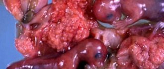
Types of hemorrhages in the eye
Hemorrhages in the eye are divided depending on the place where the blood accumulates:
- With hyposphagma, blood accumulates between the conjunctiva and sclera due to damage to the conjunctival vessels. It looks like a bright red spot on the white of the eye. This is the least dangerous type of hemorrhage and does not lead to visual impairment, but you should be wary if it occurs regularly;
- With hyphema, blood flows into the anterior chamber of the eye. Hyphema has 4 degrees: 1st degree is characterized by filling the chamber by no more than 1/3, 2nd – by no more than 1/2, 3rd – by more than 1/2, but not completely, 4th – complete filling of the anterior chamber of the eye with blood. If blood reaches the pupil, the vision becomes blurred, vision decreases, and fear of bright light appears. With this type of hemorrhage, pain occurs;
- with hemophthalmia, blood fills the vitreous body. There are complete and incomplete hemophthalmos. Vision drops sharply, the degree of impairment of which depends on the volume of blood shed. A very dangerous type of hemorrhage that can lead to filling of the vitreous with connective tissue, permanent vision loss and possible retinal detachment;
- In retinal hemorrhages, blood accumulates just in front of the retina (preretinal hemorrhages), inside the retina (intraretinal hemorrhages), or just after the rod and cone layer (subretinal hemorrhages).
This is the most serious type of hemorrhage, most often leading to decreased or even loss of vision.
Possible causes of hemorrhage in the eye
The most likely causes of hemorrhage in the eye include:
- mechanical impacts, eye injuries, head injuries - the degree of damage depends on the force of the impact;
- intraocular operations;
- rupture of newly formed vessels;
- diseases and conditions that affect the composition and properties of blood, disrupting the blood clotting process (taking blood-thinning drugs, hematological diseases);
- diseases affecting the vascular wall (hypertension, atherosclerosis, diabetes mellitus);
- eye diseases: glaucoma (with increased intraocular pressure), high degree of myopia, retinal detachment, thrombosis (blockage) of the central retinal vein;
- various infections (viral, bacterial, fungal, etc.) leading to inflammation of the eye structures. Conjunctivitis (inflammation of the conjunctiva), iritis (irises), iridocyclitis (iris and ciliary body), chorioretinitis (choroid and retina), retinitis (retina only);
- increased intracranial pressure due to excess volume in the cranial cavity due to intracranial hematomas, brain tumors, cerebral edema;
- an increase in intra-abdominal and, as a consequence, venous pressure during severe physical exertion, debilitating vomiting, and during childbirth.
For non-penetrating blunt eye injuries
The cause of vascular rupture is a sharp increase in intraocular pressure, the same mechanism is triggered in glaucoma.
Newly formed vessels have a rather fragile wall; they can appear without good reason or to replace damaged ones, for example, in diabetes mellitus
. In addition, an increase in blood sugar levels leads to damage to the smallest vessels - capillaries.
For infectious diseases
the inflammatory process leads to increased vascular permeability, which can cause damage.
Blood diseases that most often lead to hemorrhages include hemorrhagic diathesis
.
This is a group of diseases in which the processes of stopping bleeding are disrupted.
For example, in hemophilia, there is a hereditary decrease in blood clotting factors, which prevents a blood clot from forming. In thrombocytopenia, there are not enough platelets to initially block the lesion. Oncological diseases of the blood. The most striking example is acute leukemia
. Tumor cells displace bone marrow cells, then enter the bloodstream and replace all healthy cells, including those responsible for stopping bleeding.
Atherosclerosis
manifested by the deposition of cholesterol plaques in blood vessels.
A constant or periodic increase in blood pressure
damages the inner layer of blood vessels - the endothelium, while the middle, muscular layer thickens, and the vessel loses its elasticity. These changes sooner or later lead to a violation of the integrity of any vessel.
Retinal disinsertion
from the choroid with hemorrhage occurs with eye injuries, inflammatory diseases of the retina, and disturbances in its nutrition. The risk of detachment increases in people with a high degree of myopia.
Symptoms of retinal detachment include a sharp decrease in vision, narrowing of the visual field or loss of a portion of the image, the presence of spots in front of the eyes, flashes of light.
Which doctors should you contact if there is a hemorrhage in the eye?
The treatment of hemorrhages in the eye is carried out by an ophthalmologist; to correct concomitant conditions, a consultation with a general practitioner may be required.
Diagnosis and examinations for hemorrhage in the eye
The doctor conducts a survey and examination of the patient and his eyes, determines visual acuity, measures blood pressure, intraocular pressure, studies the structures of the anterior chamber of the eye, and examines the fundus, if possible.
A number of additional examination methods may be required:
- ultrasound examination of the eye;
- electroretinography to assess the functional activity of the retina;
- general blood analysis;
How to help your pet
Helping your pet can go in two directions: eliminating the cause and symptomatic relief of the condition. In order to eliminate the cause, in most cases a consultation with a veterinarian is necessary.
At home, you can carry out hygiene procedures and use eye drops. For rinsing, it is better to use saline solution (at a concentration of 0.9%) or special drops. At home, non-concentrated herbal decoctions (for example, chamomile) can be used for preventive rinsing. You need to rinse your eyes at least 2 times a day (morning and evening).