Almost every veterinary specialist knows that an animal’s eyes are a “mirror” by which a specialist can visually determine if the animal is sick or not. Today in medicine, many human diseases are diagnosed by the eyes.
Eye diseases in our cats are quite common today. If you do not take the necessary treatment measures in a timely manner, sometimes your pet’s disease can result in loss of vision.
Before talking about eye diseases, cat owners should have an idea of what the eye is.
A cat's eye is an organ specially adapted for the animal to perceive light waves. With the help of vision, your cat navigates the world around it, perceives the intensity of light, color, shape of objects, distance to them, as well as the movement of objects in space. The eyeball of a cat has a shape close to a ball and consists of an optical part and a photoreceptor part. Light rays, before reaching photoreceptor cells, pass through a complex optical system of transparent media. The first thing the rays encounter on their way is the cornea, part of the white outer shell - the sclera. Externally, the cornea resembles a slightly convex watch glass; Unlike the sclera, the cornea is devoid of blood vessels and is almost transparent. A ray of light passing through the cornea enters the anterior chamber of the eye (the space between the cornea and the lens). This space is filled with liquid (chamber moisture). Adjacent to the inner part of the sclera is the second layer of the eye - the vascular layer, which is rich in arterial and venous vessels. In the front part of the eye, the choroid turns into the iris, which contains the pigment that gives the eye its color (yellow, green, sometimes blue).
The iris, just like the aperture in a camera, regulates the amount of light entering the eye. The iris is equipped with two layers of ciliary muscles: annular and radial. In the middle of the iris there is a hole - the pupil, through which a ray of light enters the back of the eye. If a cat is in the dark and is suddenly illuminated, then as a result of contraction of the annular muscle, the pupil narrows and the flow of rays entering the eye decreases.
After passing through the pupil, a ray of light enters the lens - a transparent body similar to a small, biconvex magnifying glass. Along the edge of the entire circumference of the lens, the ligament of zinn is attached to it. The lens itself is enclosed in a capsule and attached to the ciliary muscle through the ligament of cinnamon.
From the lens, the light beam passes through the vitreous body of the eye, which mainly fills the eyeball. The vitreous body is transparent and consists of the finest fibers, which are a skeleton, between which there is liquid.
The cornea, aqueous humor of the anterior chamber, lens and vitreous body constitute the optical, or refractive, system of the eye. Before affecting the retina, the light beam changes its direction many times, due to the fact that each of these media has its own refractive index.
Adjacent to the vitreous body is the third layer of the eye, the retina. Its outer layer consists of pigment cells that prevent the scattering of light rays, while the inner layer consists of cells called rods and cones. They are the receptor, or perceptive, department of the visual analyzer.
When examined with an ophthalmoscope, the back wall of the eyeball, the so-called fundus, reveals a pale colored area from which blood vessels radiate. Experts call this area a blind spot because there are no light-sensitive cells in it. From the entire retina, nerve fibers converge to the blind spot to form the optic nerve. The total number of light-sensitive cells in a cat's eye is very large. At the same time, rods, unlike cones, have a higher sensitivity to light.
Under the influence of light, the metabolism of the retinal pigment cells changes and electrical phenomena occur. The reddish pigment rhodopsin, or visual purple, contained in the rods, turns into the yellow pigment retinen in the light. In the dark the reverse transformation occurs. Both of these pigments consist of protein and vitamin A. The quantitative ratios of rhodopsin, retinene and vitamin A are established depending on the degree of illumination of the retina.
Today, all existing eye diseases in cats are divided by experts into two large groups:
- Diseases and damage to the protective devices of the visual organs.
- Diseases and damage directly to the eyeball itself.
Veterinary specialists include diseases and damage to the protective devices of the visual organs:
- Bruises and other mechanical damage in which the integrity of the skin is not damaged.
- Mechanical injuries and wounds accompanied by skin damage and bleeding.
- Inflammation of the eyelids (blepharitis).
- Turning of the eyelids.
- Eversion of the eyelids.
- Inability to close the eyelids (lagophthalmos).
- Upper eyelid incontinence (ptosis).
- Tumors and various neoplasms.
Diseases and injuries of the eyeball include:
- Prolapse of the third eyelid.
- Conjunctivitis.
- Blepharitis (inflammation of the eyelids).
- Keratitis (inflammation of the cornea).
- Corneal ulcer.
- Cataract (clouding of the lens).
- Glaucoma (increased intraocular pressure).
- Iritis or iridocyclitis (disease of the ciliary body and iris).
- Dacryocystitis (impaired patency of the lacrimal canal).
- Exophthalmos (prolapse of the eyeball).
- Enophthalmos (sinking of the eyeball inwards).
- Panophthalmitis (inflammation of all membranes of the eye).
All of the above eye diseases can be of infectious or non-infectious etiology.
Eye diseases in cats are characterized by the presence of similar symptoms of the disease:
- lacrimation.
- redness and swelling of the eyelids and conjunctiva.
- various discharges from the eyes.
- soreness.
- photophobia.
- Sometimes in the morning the cat is unable to open the affected eye.
- itching appears in the affected eye.
Diagnosis of eye diseases.
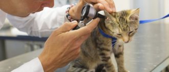
Diagnosis of a particular eye disease in cats should be carried out by specialists from veterinary clinics. At the veterinary clinic, specialists will conduct a clinical examination of your cat to rule out the presence of infectious diseases that have led to damage to the visual organs. The affected eye is examined more deeply (the size and shape of the pupils, the size of the palpebral fissure, the size of the eyeballs, the presence of injuries, etc.). If necessary, smears will be taken for culture on nutrient media. Blood will be taken to rule out infectious diseases.
Diseases of the eyes and protective devices of the visual organs
Bruises and mechanical damage without compromising the integrity of the skin.
Cause. They occur in a cat as a result of a blow with a blunt object or as a result of mechanical damage (bumping into blunt objects, falling).
Clinical signs. During a clinical examination, the veterinarian notes the presence of bruises and hematomas in the eye area. In the area of injury, upon palpation, an increase in local temperature is noted, and a change in the size of the eyeball occurs.
Treatment. A cat with an eye injury should be taken to a veterinary clinic. To relieve pain in a sick animal, a 2% solution of novocaine, antimicrobial drops or eye ointment is instilled into the conjunctival sac. If there is a hematoma, it is necessary to apply cold to the hematoma (only on the eyelid area, not on the eyeball). If there are serious complications, it is sometimes necessary to resort to surgery.
Diagnostics
It is carried out by a veterinarian in a specialized clinic, as it requires special tools. It is carried out in several stages. First, the organ is examined and the following parameters are assessed:
- preservation of vision;
- appearance of the organ: the size and shape of the pupils, their symmetry; the same parameters are used to evaluate the eyelids and their condition;
- condition of the eye: its size, shape, position relative to the orbit, absence or presence of injuries.
After an external examination of the organ, the doctor begins research using additional equipment: examines the fundus of the animal, measures intraocular pressure, prescribes general clinical and biochemical tests of blood and urine, takes a smear to identify pathogenic microflora and its sensitivity to antibiotics, etc.
Wounds and open injuries to the eye.
Cause. Most eye injuries and wounds in cats occur as a result of fights with their fellow cats, falling on sharp objects, or strong blows leading to damage to the skin.
Clinical signs. During a clinical examination, the veterinarian notes a violation of the skin in the area of the injured eye, the presence of scratches, wounds, and bleeding from the injured areas.
Treatment. The damaged area is washed with hydrogen peroxide, bleeding is stopped; in the presence of large wounds, pain relief and suturing are used, and antimicrobial therapy is administered locally. For complex and large eye injuries, eye microsurgery and sometimes removal of the damaged eye are performed.
Eye diseases of cats
Diseases affecting the eyelids of an animal include:
- Inflammation of the eyelid (blepharitis).
- Wounds and bruises.
- Entropion or inversion of the eyelid.
- Ptosis (drooping of the upper eyelids).
- Lagophthalmos (fusion) of the eyelids.
- Neoplasms.
affect the eyeball :
- Conjunctivitis.
- Glaucoma (high blood pressure).
- Cataract.
- Dermoid (neoplasm in the conjunctiva).
- Dislocation of the eyeball.
- Corneal ulcers and inflammation.
- Keratitis.
Blepharitis
Blepharitis is inflammation of the eyelids.
Cause. Traumatic damage to the eyelids, the presence of a bacterial or fungal infection, allergies in animals, diabetes (diabetes mellitus in animals, diabetes insipidus in animals), liver disease.
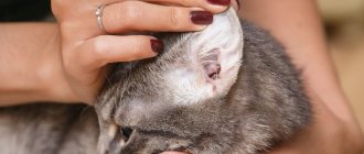
Clinical signs. During a clinical examination, a veterinarian notes redness of the eyelids, swelling, photophobia, profuse lacrimation, itching and burning in the area of inflammation of the eyelids, loss of hair and eyelashes around the eyelids.
Treatment of blepharitis should be aimed at eliminating the cause that led to blepharitis. The sore eye is washed with a decoction of calendula or chamomile, a solution of boric acid, potassium permanganate or furacillin. Eye drops are instilled into the conjunctival sac. If infection is present, antibiotics are used.
Prevention
The owner of the animal must be attentive to his well-being and behavior, and notice any changes in this.
If a cat rubs its eyes and shows anxiety, it is worth examining it, the cause may be a foreign body. If this is the case, you need to remove it and rinse the eye with plenty of clean water, furatsilin, a weak solution of potassium permanganate, and then apply any eye drops. After this, you need to take your pet to a veterinarian for a more thorough examination.
For preventive purposes, it is worth wiping your pet’s eyes daily with boiled water or chamomile decoction. Animals do not really like hygiene procedures, and therefore it is necessary to accustom them to this from childhood.
If crusts, discharge, or any changes in the pet’s behavior are detected, it is necessary to show it to a veterinarian as soon as possible.
1111
Entropion of the eyelids
Cause. Entropion of the eyelids in cats occurs as a result of certain eye diseases (conjunctivitis, blepharitis, etc.) and exposure to certain chemicals. A genetic predisposition in some cat breeds (Sphynxes, Persians) leads to bloat.
Clinical signs. During a clinical examination, the veterinarian notes that the eyelid turns inward. As a result of mechanical irritation, redness of the eyeball and symptoms of blepharospasm are noted. When examining the cornea, we record the presence of ulcers in the place of constant contact with the eyelid, and lacrimation.
Treatment. Carrying out a surgical operation.
Inflammation of the nasolacrimal ducts
Epiphora is a disease of the nasolacrimal ducts, which is expressed in their blockage and inflammation.
The reasons lie in:
- breed predisposition (Scottish, Persian, exotic cats);
- congenital absence or anatomical narrowness of the neck and mouth of the nasolacrimal ducts;
- injury;
- viral, bacterial, chlamydial infections;
- dacryocystitis;
- canaliculitis.
Inflammation of the nasolacrimal ducts can be diagnosed using a fluorescein test. Before it is performed, the eyes are washed with an antibiotic solution, removing all purulent discharge. Then 1-2 drops of a special composition are instilled into the eye, and the cat’s head is tilted forward. After 2 minutes, the oral cavity of a healthy animal will turn green. If there is partial blockage, coloring will occur within 5-10 minutes. In case of complete obstruction, the natural shade of the mucous membrane will remain unchanged.
Treatment of inflammation of the nasolacrimal ducts is carried out with the help of antibacterial agents, antiseptics and antihistamines. If a cat has a complete blockage of the canals, the veterinarian may resort to surgical bougienage followed by eye hygiene.
Wounds of the eyelids
Wounds to the eyelids are most common in cats that are kept free-range and interact with their cats. Fights for territory and hierarchical fights often end in deep wounds, complicated by infections and street dirt. Untreated wounds in the eye area are accompanied by swelling, hyperemia, and suppuration.
They can be superficial, deep and through. It is enough to clean the superficial wound from dirt and treat it with an antiseptic composition. A deep wound can be recognized by uneven, swollen edges, extensive tissue tears, and copious discharge in the form of ichor or pus. Self-medication of such injuries leads to scar formation and loss of vision.
The veterinarian should examine the patient and determine the extent of tissue damage. Only after this can you choose an effective scheme for restoring cat eyes.
Turn of the century
Entropion (or entropion) is one of the most common ophthalmic diseases in cats. It lies in the abnormal position of the eyelid relative to other structures of the eye.
Entropion can be:
- one-sided or two-sided;
- bottom or top.
The development of pathology is due to the weakness of the connective tissue on the inner lining of the eyelid.
Risk factors include:
- panophthalmitis, uveitis and other inflammations;
- injuries;
- microblepharon (an animal’s inability to close its eyelids tightly, which in winter leads to thermal damage to the cornea);
- corneal syndrome (formation of ulcers on the cornea with copious purulent discharge);
- age-related loss of muscle tone;
- oncological neoplasms.
In the initial stages, the disease manifests itself in frequent blinking, photophobia, and severe pain. As the pathology develops, the discharge becomes cloudy, strabismus appears, the eyelids swell, and the body temperature rises. Transition to the chronic form threatens the cat with complete blindness.
In some cases, conservative treatment is acceptable, but most often doctors recommend surgery.
Glaucoma
Glaucoma is a disease associated with atrophy of the optic nerve and a regular increase in intraocular pressure.
The most common causes of glaucoma are:
- trauma and inflammation of the lens;
- getting particles into the eye;
- chronic purulent inflammation of the eyes;
- changes in the cornea.
The main symptoms of the disease are:
- enlargement and hardening of the eyeball;
- pupil enlargement;
- swelling of the mucous membranes;
- cloudiness and loss of sensitivity of the cornea;
- dark red eye color.
The initial stages of glaucoma are treated with conservative methods using diuretics and antibacterial drugs. When the disease is advanced, cats are operated on.
Eversion of the eyelids
Cause. Eversion occurs as a result of certain eye diseases, when they become chronic. Eversion of the eyelids is common in some cat breeds. It is quite rare in clinical practice.
Clinical signs. During a clinical examination of a sick cat, a veterinarian notes blepharospasm (spastic closure of the eye), conjunctivitis, lacrimation, and at the site of ectropion the veterinarian diagnoses the area of the mucous membrane.
Treatment. Surgical intervention.
Obstruction of the lacrimal duct
Tears must flow through a special channel that is located inside the nose, but if the outflow path is disrupted, the tears come out through the eyes. The main symptom is different types of lacrimation and the appearance of brown fur around the eyes (tear tracks). A similar problem can be congenital (found in some breeds - Sphynx, Persian or Scottish cats) or acquired (obstruction can be caused by various infections).
If patency occurs due to blockage or fusion of the nasolacrimal duct, surgical intervention may be prescribed. Although the use of special rinses and preparations is usually sufficient.
Fusion of eyelids
Cause. Eyelid fusion in a cat can be either congenital or acquired. Owners notice congenital fusion of the eyelids after the birth of a kitten. Acquired fusion in a cat occurs after injuries to the head and eyes, with chronic blepharitis, chemical and thermal burns of the eyelids.
Clinical picture. During a clinical examination, the veterinarian cannot separate the eyelids, there is a continuous strip of skin between the eyelids, and there is a scar between the fused eyelids.
Treatment. Surgical intervention aimed at separating the eyelids.
When veterinary help is urgently needed
If something is wrong with a cat’s eye, the owner can provide first aid. After this, it is recommended to consult a doctor. This must be done even if the problem seems completely insignificant. After all, it is impossible to make a diagnosis on your own, and your mistake can result in premature blindness of your beloved pet.
Emergency assistance will be required if pus appears, high fever, signs of cataracts and glaucoma, burns or other health-threatening injuries. In these cases, it is recommended to contact the nearest 24-hour veterinary clinic without waiting until morning.
Lagophthalmos
Lagophthalmos in a cat is manifested by the inability to completely cover the eyes with the eyelids.
Cause. Eversion and inversion of the eyelids, paralysis of the facial eyelid, hereditary shortness of the eyelids.
Clinical signs. During a clinical examination of a sick cat, a veterinarian notes a constantly slightly open palpebral fissure, lacrimation, and lag of the lower eyelid from the eyeball.
Treatment. Treatment of this pathology in cats is surgical, and it is necessary to first use antimicrobial eye drops and corneal protector medications.
Classification
There are several of them. By type of process there are:
- Inflammatory. These include conjunctivitis, keratitis, keratoconjunctivitis, iritis (the iris is damaged), inflammation of the lacrimal canal, etc.
- Not inflammatory. Injuries, bruises, foreign bodies entering the eye, entropion of the eyelids, cataracts, glaucoma, etc.
According to the course of the process, the disease can be acute, subacute and chronic. In the subacute process, the symptoms do not disappear completely, but only subside slightly, while the danger of vision deterioration remains. This course is no less dangerous than acute or chronic, although it seems that the animal has gotten better.
They are divided into:
- Primary – eye problem – this is the underlying disease.
- Secondary - the organ of vision reacted in response to concomitant pathology. For example, when a cat is infected with bacteria, in addition to other organs, the eyes are also affected, which manifests itself in the form of conjunctivitis. In this case, it is necessary to identify the disease that caused this condition and treat the animal comprehensively, and not just deal with the symptoms.
Uevit
Uevitis is an inflammation of the choroid of the eye.
Cause. Uevitis in cats occurs as a result of eye injuries, metabolic disorders, eye tumors, autoimmune diseases, toxoplasmosis, rickettsiosis, diseases caused by viruses (feline leukemia, feline immunodeficiency, infectious peritonitis, etc.).
Clinic. Uevitis in a cat is accompanied by photophobia, lacrimation, eye pain, blepharospasm, strabismus, the cat's pupil is greatly reduced or takes on an uneven, blurry shape. The eyes have a red and cloudy appearance on clinical examination. There is a change in the color of the iris.
Treatment of ueviitis in a cat should be aimed at the cause that led to the disease. Eye drops are instilled into the eyes and painkillers are prescribed. Antibiotics and other antimicrobial drugs are used. If there is a threat of developing glaucoma, it is necessary to resort to surgical removal of the diseased eye.
Symptoms of diseases
Among the most common are the following diseases.
Conjunctivitis
It is characterized by the presence of an inflammatory process on the mucous membrane of the eyelids. One of the most common in cats.
There are several types: catarrhal, purulent, ulcerative, follicular. It occurs as a result of various reasons: foreign bodies, vitamin deficiency, injuries, as a consequence of many infectious processes. For example, a common runny nose.
This pathology is manifested by redness of one or both eyelids, their swelling and lacrimation. This picture is typical for catarrhal type; with purulent form, the symptoms are the same, but more pronounced and, in addition to the copious secretion of tear fluid, purulent discharge appears. The swelling can be so severe that the eyelids hardly open, and they also stick together, making it difficult for the cat to open them.
When the process is advanced or immunity is reduced, a rise in local or general temperature in the animal is possible. With follicular conjunctivitis, in addition to the described symptoms, regional lymph nodes located in the inner corner of the eyes are affected.
Cataract
Characterized by clouding of the lens. Older animals are more susceptible to this disease, but young animals can also develop it as a result of infection.
Main symptom: decreased vision. The pet becomes slower and more careful, loses orientation in an unknown environment, and crashes into everything.
Dacryocystitis
Inflammation of the lacrimal sac. It is divided into acquired and congenital. This disease develops as a complication after others, such as injuries or infections.
All age categories of pets are susceptible to it.
The main symptoms are: swelling of the conjunctiva, lacrimation, redness in the corners of the eyes, the lacrimal sac increases in size, temperature rise, and pus is released when pressed. The cat becomes restless due to severe pain that develops with this disease.
Manifestation of keratitis
Keratitis
This is an inflammation of the cornea. It occurs as a result of other pathologies: conjunctivitis, thermal burns, infections, allergic reactions, vitamin deficiency, injuries and congenital predisposition.
Some cat breeds are most susceptible to keratitis, for example, British, Siamese and Persian, American Smooth-haired.
It is characterized by clouding of the cornea and swelling of the upper eyelid. Blood vessels become noticeable as red streaks, and pus accumulates in the corners of the eyes. The cat stops liking the light, begins to squint and hide in dark places.
An ulcer of the cornea of the eye occurs for a number of reasons: chemical burns with cleaning and disinfection products, ingrown eyelashes, trauma, damage by viruses and bacteria. Breeds with flat faces and protruding eyes are more susceptible.
Symptoms: the organs of vision become red and as if “cloudy”, copious discharge, the pet constantly scratches his eyelids, trying to relieve the itching and squints, bright light becomes unpleasant for him.
Third eyelid (loss)
This is a nictitating membrane, which is a protective device. It serves to protect the animal's eyes from mechanical damage and foreign bodies. It can be seen on a pet without special tools; it is thin, almost transparent, light skin near the inner corner of the eye. It helps in moisturizing the eyes. Present in cats, birds and some mammals.
Its loss is characterized by spasms of the eye muscles (uncontrolled closing and opening of the eyelids, their twitching), redness, lacrimation, suppuration with the formation of crusts.
The disease is similar to an adenoma, and therefore you should definitely consult a veterinarian, since if treated incorrectly, it can develop into keratoconjunctivitis. This disease cannot be completely cured and the animal will have to suffer from it for the rest of its life.
Prolapse of the nictitating membrane develops for various reasons, the main ones:
- conjunctivitis;
- eye injuries;
- diseases of internal organs and systems;
- allergic reactions;
- the presence of parasites and helminths;
- fungal skin infections.
Sometimes situations arise when the third eyelid falls out in only one of the eyes, this is often caused by debris getting under it, injuries or a unilateral bacterial infection.
If this situation is bilateral, then most likely the reason is the fusion of part of the eyelid with the eyeball, conjunctivitis or helminthic infestation.
Blepharitis
This is inflammation of the eyelid. It is divided into: simple, ulcerative, scaly, meibomian.
If you notice redness in the eyelids of your pet, you should immediately contact a veterinary clinic. This simple, at first glance, condition very quickly turns into an ulcerative form, and this is already a serious disease.
Manifestation of blepharitis
If this pathology is left untreated, the pet may even lose an eye, as the infection spreads directly to the organ of vision. Panophthalmitis develops, a disease that is often the reason for the removal of an animal’s eyes due to the lack of effectiveness of conservative treatment.
In addition to redness of the eyelid itself, a characteristic sign is the pet’s desire to scratch it. It is worth paying close attention to this behavior and not leaving everything to chance, as the cat can additionally cause an infection or injure the eye with its paw and claws.
Panophthalmitis
A rather rare, but extremely serious infectious disease, manifested by inflammation in all tissues of the eyeball.
Characterized by symptoms: an increase in the size of the organ with a large amount of purulent discharge.
If your pet has been diagnosed with this, you will have to part with the eye, as there is a risk of the infectious process spreading to the brain.
Therefore, if you notice the first signs of eye problems, you should see a veterinarian rather than self-medicate or wait for it to go away on its own. After all, this will waste precious time for treatment and preservation of the organ.
Eyelid wounds are a common occurrence in cats; they get them in fights with other animals. They are divided into:
- superficial (scratches);
- deep (several layers of skin are damaged);
- through (the eyelid is torn right through).
Manifestation of panophthalmitis in kittens
If the outer side of the eyelid is scratched, you can use home remedies: wash the wound with an antiseptic and blot with gauze. If the damage is deeper, you should see a specialist, as surgical intervention is often required to apply sutures and remove foreign particles from the wound.
Entropion of the eyelids
The lower eyelid is most susceptible to this. It bends towards the cornea and causes severe pain to the animal. In cases of repeated volvulus, surgical treatment is performed. The disease is often the cause of chronic keratoconjunctivitis.
Glaucoma
Characterized by increased intraocular pressure.
There are types:
- closed-coal;
- congenital;
- open-coal.
Among the main symptoms are: pupil dilation, burst vessels on the conjunctiva and the eye itself, an increase in the size of the organ and it becomes hard (felt when pressed lightly). Normally, the eyeball is quite elastic and, with light pressure, springs a little under the finger, but with glaucoma it becomes as if it were made of stone. This occurs due to a sharp increase in pressure inside.
Glaucoma in a cat
In the open-angle form of the disease, clouding of the cornea occurs and its sensitivity decreases. The development of glaucoma leads to decreased vision and causes suffering to the pet due to incessant pain in the eye, due to which the pet becomes restless, sleeps poorly and meows a lot. Therefore, if you notice the symptoms described above, you should urgently contact a veterinarian to prescribe adequate treatment.
Sometimes glaucoma requires urgent surgery, for example, when the lens is dislocated, which is the cause of the disease.
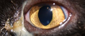
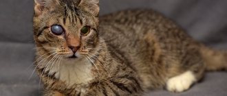
Belmo
A very common pathology among cats. It is very easy to notice - an area of turbidity or whitening appears on the cornea. It can occur as a result of injury, an infectious process, or untreated cataracts. This disease causes vision impairment in 90% of cases.
Cat's thorn
There are a number of other symptoms:
- in 50% of cases strabismus develops;
- the cornea is red, swollen;
- eyes fester;
- increased lacrimation;
- the animal has a negative attitude towards the owner’s attempt to wash his eyes.
Sometimes an eyesore leads to the development of glaucoma, in which case they resort to radical measures and remove the eye, while the eyelids are sutured.
Third eyelid prolapse
Prolapse of the third eyelid in a cat is often a secondary symptom of a number of infectious and parasitic eye diseases; it occurs when the optic nerve becomes inflamed.
Clinic. Cat owners and veterinary specialists use eyelids, which sometimes reach up to 1/3 of the entire visual area.
Treatment. Third eyelid prolapse in cats can be treated symptomatically or surgically. Treatment should be aimed at eliminating the primary disease that led to the loss of the third eyelid. Surgical treatment involves surgical excision of the prolapsed third eyelid.
Symblepharon
Symblepharon is a fusion of the conjunctiva with the eyelids and cornea.
Symptoms:
Symblepharon varies in area of damage and localization: it can be partial or complete.
If it is small in size, it is sometimes an accidental finding during a general examination, but if it is of a significant size, it is already a cosmetic defect and interferes with the cat’s vision, but the symblepharon itself does not bother the cat.
With partial symblepharon, when the conjunctiva may not fuse with the cornea, the prognosis is favorable; with complete symblepharon, the prognosis is most often unfavorable.
Treatment:
1. Daily eye sanitation.
2. Separation of the conjunctiva from adjacent layers, the cornea, using local preparations with antibiotics and steroidal anti-inflammatory drugs.
3. In case of severe symblepharon, leading to visual impairment, the option of surgical treatment to separate the fused tissues is considered.
============================================================================================================================================================================================
Keratitis
Keratitis is inflammation of the cornea.
Cause. Keratitis in cats is caused by infectious diseases (infectious rhinotracheitis in cats, feline chlamydia, mycoplasmosis in cats, calicivirus infection of cats), helminthic infestations, damage to the cornea by a foreign body, and eye injuries. Often the cause of keratitis can be inflammatory processes in nearby tissues.
Clinic. Keratitis in a cat can be superficial, deep and ulcerative. With keratitis, a cat may experience vision loss. During a clinical examination of a sick animal, a veterinarian notes redness of the eye, photophobia, clouding of the cornea, pain, and visible blood vessels. Cloudiness of the cornea occurs due to cellular infiltration (by leukocytes) and changes associated with cell swelling, their degenerative decay, as well as changes in the intercellular connective tissue. The intensity of turbidity can be different and depends on the density and nature of the infiltrate or edema in the interstitial tissue. The gray-smoky color of the cloudiness is formed by a small accumulation of leukocytes. With an increase in the number of leukocytes, the color of the cornea becomes white; a yellowish tint characterizes a purulent infiltrate. The opacification may be diffuse or limited to isolated areas in the form of dots or spots in the anterior layers (superficial keratitis) in the parenchyma (parenchymal keratitis). If the surface layers are damaged, the cornea loses its specular properties and becomes matte. With keratitis, vascularization of the cornea also occurs (ingrowth of blood vessels into the cornea). At the same time, during a clinical examination, the veterinarian notes a pericorneal injection, which is characterized by overflow of densely located vessels in the limbus area. The reaction from the conjunctiva, mainly its scleral part, is manifested by edema and hyperemia. With keratitis, experts note a reaction from the iris. The membrane becomes hyperemic, the pupil narrows, and serous, fibrinous or purulent exudate sometimes appears in the anterior chamber. With acute keratitis, a cat experiences photophobia, spasm of the eyelids, lacrimation and pain. In eosinophilic keratitis, veterinarians upon clinical examination detect the presence of white plaques on the cornea that extend from behind to the anterior wall. Slight lacrimation is noted. When performing a cytological examination, we find a large number of eosinophils.
Treatment. Symptomatic treatment is carried out. Eliminate the underlying disease that caused the keratitis. The affected eye is washed with antimicrobial solutions, and eye ointments and drops are used. If a purulent process occurs, antibiotics are used; for deep and ulcerative keratitis, eye microsurgery is used. For eosinophilic keratitis, they resort to the use of immunomodulators and hormones.
Conjunctivitis
This disease is known to everyone - it is one of the most common. Conjunctivitis is an inflammation of the conjunctiva, the mucous membrane of the eyelids. It is often a symptom of other global pet health problems.
Symptoms
In the photo of sick animals you can clearly see which symptoms of conjunctivitis are most active. These include watery eyes, swelling and redness of the eyelids, and noticeable clouding of the eye.
Causes
Many factors can cause inflammation of the conjunctiva. This is why conjunctivitis is so common. The main reasons for its appearance include:
- fungi, bacteria and viruses. They can affect only the membrane of the eyelid, or they can affect the entire body of the animal;
- damage and injury that violate the integrity of the protective membranes, thereby allowing infection to pass through;
- allergens (in this case, conjunctivitis is just one of the symptoms. Itching, runny nose, and sneezing may also occur);
- colds, weakened immunity;
- parasites;
- ultraviolet radiation.
Types of conjunctivitis
Based on the manifestation and source of inflammation, conjunctivitis is divided into four types that can transform into each other.
- Catarrhal . It is easily treated if measures are taken in time. Characterized by swelling, lacrimation and mucous discharge. Possible eversion of the eyelid. If you do not fight the disease, it goes into the next form.
- Purulent . Identified by the discharge of pus. Depending on the severity, it can be just yellow crusts in the morning, or obvious yellow-green pus. The eyelids may stick together. Serious complications and loss of vision are possible if you do not contact a veterinarian promptly.
- Phlegmonous . With this type of conjunctivitis, pus accumulates under the skin, so independent treatment is impossible.
- Follicular . This type is chronic. With it, not only the conjunctiva becomes inflamed, but also the inside of the third eyelid. Treatment is often impossible without surgery.
The first form can be cured on your own. Tetracycline ointment 1%, which can be easily purchased at any pharmacy, will help with this. Use a cotton swab to apply it to the lower eyelid of the animal. Repeat the procedure several times a day.
If treatment does not bring results, or a purulent form of conjunctivitis is possible, you should immediately consult a veterinarian.
Corneal ulcer
A corneal ulcer in a cat is accompanied by tissue destruction and poor healing. Superficial disintegration of the epithelium is called erosion.
Cause. The causes of the development of corneal ulcers can be both exogenous and endogenous. Exogenous causes include various traumatic injuries caused by the entry of hard and uneven foreign bodies into the conjunctival sac or as a result of direct injury to the cornea. Ulcers in cats occur when the eyelids or eyelashes roll up, when the cornea is constantly subjected to friction by the rolled-up edge of the eyelids or eyelashes. Chemical and thermal burns are always accompanied by an ulcerative process in a cat. Purulent conjunctivitis can cause the formation of corneal ulcers. Corneal ulcers can be observed when there is a violation of its innervation (neuroparalytic keratitis). Endogenous causes of ulcers in cats include a number of infectious diseases of cats, metabolic diseases, hypovitaminosis diseases, etc.
Clinical picture. Large ulcers are detected by a veterinary specialist at the clinic during examination - a defect, clouding of the cornea and discharged exudate are revealed. Small and superficial ulcers are determined by a veterinarian using side lighting and clouding of the cornea. Ulcerative processes in cats are in all cases accompanied by spasm of the eyelids, their swelling, mucopurulent, and more often purulent discharge, and the cat’s body temperature rises.
After the ulcers heal, scars form.
Treatment. Treatment of corneal ulcers is symptomatic or surgical. In the case when the eyeball is dissolved by a purulent process, it is removed. In veterinary clinics, sick animals are given antimicrobial therapy, including antibiotics, eye drops and ointments, and pain relief (novocaine blockades). Eye microsurgery is also performed to remove damaged areas of the cornea.
Other pathologies
Less common eye diseases in cats include the following:
:
- Canaliculitis
, or inflammation of the tear ducts. Occurs when the ducts become clogged and is accompanied by profuse lacrimation. It is treated by washing the clogged lacrimal sac or removing it.
- Exophthalmos (proptosis) and enophthalmos
, or prolapse and retraction of the eyeball. Changes are visible to the naked eye. The organ is returned to its original position under general anesthesia.
- Panophthalmitis
. A most dangerous pathology that affects all eye tissues. Accompanied by an enlargement of the eye and the discharge of pus. To avoid brain infection, the affected organ is removed.
- Adenoma of the third century
. It is characterized by severe redness and thickening of the third eyelid as a result of inflammation of the lymph nodes. This makes it look like a cherry. The organ affected by the adenoma is sutured to its normal state or removed.
Due to the high likelihood of complications, it is better to entrust treatment to a veterinarian. Self-administration of drugs rarely brings the desired result. At best, you will simply smooth out the existing symptoms and complicate the diagnosis.
Iritis (iridocyclitis)
Iritis (iridocyclitis) is inflammation of the iris and ciliary body.
Cause. Traumatic injury, inflammation spreading from the cornea, complications after eye surgery, infectious diseases.
Clinic. During a clinical examination, a clinic veterinarian notes a constriction of the pupil, dimming of the iris, photophobia, pain, and cloudy fluid in the anterior chamber of a sick cat. When conjunctivitis is associated with the disease, we note serous-purulent discharge.
Treatment. In order to restore pupil contraction, a 1% atropine solution, novocaine blockade, and antibiotics are prescribed. During treatment, eye drops and ointments with anti-inflammatory and antimicrobial effects have a good effect.
Treatment at home
After the diagnosis is made, the mustachioed patient must be provided with the most comfortable conditions possible. Place it in a darkened room, away from noise sources, other pets, children and other irritants.
To avoid further injuries, be sure to trim your cat’s claws and do not let him outside until he recovers. In addition to the basic treatment, which includes the use of eye drops and ointments, you will need to pay close attention to hygiene through rinsing.
Washing and hygiene
Washing is carried out using cotton pads, guided by the following algorithm
:
- Prepare all the necessary tools and preparations in advance to reduce the risk of traumatizing the animal with painful waiting.
- Wrap your pet in a thick cloth or find an assistant who can hold him during the procedure.
- Soak a cotton pad in the medicinal solution, squeeze it out and open the cat's eyelids.
- Gently move the disc over the eye, moving from the outer corner to the inner.
- Repeat the previous steps with the other eye, not forgetting to change the cotton pad.
The procedure can also be carried out using a syringe without a needle. To do this, you need to irrigate each eye with the solution under low pressure and gently blot them with a clean, lint-free cloth. After removing the accumulated dirt and soaking the crusts, you can begin applying the main preparation.
Drops and ointments
If something is wrong with a cat’s eye, then special drops and ointments will be required to normalize its condition. The list of necessary medications will depend on the cause of the disease:
- bacterial infection – Iris, Leopard, Levomycetin;
- viral infection – Anandin;
- fungal infection – Tetracycline;
- injuries - Traumeel.
The drops are instilled into both eyes at once, and the ointment is applied only to the disturbing area. After application, close the eyelids tightly and gently massage so that the product is evenly distributed inside. During treatment, it is recommended to wear an Elizabethan collar on your pet so that it does not inflict further injuries on itself when scratching.
Is it possible to use folk remedies?
Folk remedies can only be used as part of complex therapy. They do not fight the root cause, but only alleviate the patient’s condition by eliminating associated symptoms. It is also important to note that some herbs can cause an allergic reaction, so it is best to use them only under the supervision of a veterinarian.
Please note that the popular recipe with olive oil is extremely dangerous. Any oily substances create a very dense film. Because of this, new bacteria can quickly grow on the mucous membrane after treatment.
Features of treatment of pregnant, lactating and young individuals
Kittens, as well as pregnant and lactating animals, need gentler medications and lower dosages. Their immunity is weakened, so most antibiotics cause severe complications. Some of them can lead to miscarriages or stillbirths.
They also resort to surgery with extreme caution, since the anesthesia used can affect the course of pregnancy or the baby’s fragile heart. The indication for its implementation is a serious threat to life that exceeds the existing risks.
Glaucoma
Glaucoma is an increase in intraocular pressure, which is accompanied by an enlargement of the eyeball in a cat.
Glaucoma in cats is usually not a primary disease, but often accompanies other eye diseases. If treatment is not started promptly, glaucoma causes vision loss in cats. Cats are usually asymptomatic. Glaucoma can be open-angle or closed-angle.
Clinic. Angle-closure glaucoma is characterized by redness of the eye, swelling of the eyelids, severe pain, clouding of the cornea, weakness, nausea, and vomiting.
Treatment. To normalize intraocular pressure, pilocarpine is used, to improve the outflow of excess fluid in the cat’s body, diuretics, retrobulbar administration of aminazine, and the prescription of painkillers. Sometimes it is necessary to resort to surgical intervention (extirpation of the eyeball).
Third eyelid
Normally, the nictitating membrane (nictitating eyelid) performs an important protective function.
However, with some pathologies, the nictitating membrane may fall out and the so-called “third eyelid” may form in the cat. It begins to form in the corner of the eye and looks like a white film. In the normal state, it is visible only when the animal blinks or tilts its head. If you see this white film in other cases, a visit to the veterinarian is necessary.
Third eyelid prolapse is rarely an independent disease. Most often, this is a sign of other, more serious, pathological processes in the pet’s body.
Symptoms of eyelid loss are:
- redness of the conjunctiva;
- swelling;
- secretion of mucus, pus;
- lacrimation;
- formations in the corner of the eyes (the third eyelid itself);
- itching and fever are possible.
During treatment, medications are prescribed that relieve symptoms and also eliminate the cause of the disease.
Cataract
Cataract is clouding of the lens. The disease often leads the cat to blindness. Cataracts in cats are associated with metabolic disorders in the lens and are accompanied by increased breakdown of protein components.
Cause. Trauma, infectious diseases, and chronic inflammatory eye diseases lead to cataracts in cats.
Clinic. At the last stage of the disease, the cat has difficulty seeing in the affected eye. Upon examination, the lens has a white or bluish color. The dilated pupil does not respond to light.
Treatment. The use of eye drops for cataracts can only slow down the degenerative processes developing in the lens.
Causes of eye problems in cats
Three groups of reasons are to blame for the occurrence of ophthalmic diseases. These include:
- infectious, due to the negative effects of viruses and bacteria;
- non-infectious, including tumors, injuries and allergies;
- congenital, associated with breed predisposition and poor genetics.
The risk group includes extreme-type Persians with pronounced breed traits. Due to increased tear production, which forms pathogenic microflora, their tear ducts are very often infected with bacteria.
Dacryocystitis
Dacryocystitis is an inflammation of the lacrimal sac, accompanied by a violation of the patency of the nasolacrimal canal due to its narrowing.
Cause. The disease in cats occurs as a result of chronic conjunctivitis.
Clinic. During a clinical examination, a veterinarian notes mucous or purulent discharge from the eyes of a sick cat. In the outer upper corner of the eye orbit, a swelling is visible, which is painful on palpation. The upper eyelid is swollen, the lacrimal gland is enlarged on palpation. Sometimes there is an accumulation of pus in the tissues around the eyes.
Treatment. Treatment of dacryocystitis in cats can be symptomatic or surgical. Symptomatic treatment consists of relieving inflammation and pain by using novocaine blockades and absorbable ointments (Vishnevsky ointment, ichthyol). Surgical treatment consists of cleaning the lacrimal canal or extirpation of the lacrimal gland.
Blepharitis: causes, manifestations and treatment methods
Outdoor and indoor cats are susceptible to inflammation of the eyelids. This disease is called blepharitis. The pathology is rapidly worsening. In the absence of timely treatment, blepharitis turns into panophthalmitis. The advanced stage of the disease often causes the loss of one or both eyes.
Veterinarians identify the following causes of blepharitis:
- staphylococcal or streptococcal infections;
- seborrhea;
- fungal lichen;
- subcutaneous mites;
- helminths;
- endocrine disorders;
- autoimmune diseases;
- allergy;
- eye wounds;
- salivary gland duct transplantation.
Symptoms and treatment are closely related. Therapy is prescribed taking into account the type and manifestations of the pathology. Blepharitis occurs:
- Scaly. This is the most common type of illness. The cat's eyelids turn red, swell and become dense. Purulent scales form near the eyelashes. Later the hairs fall out.
- Ulcerative. This form of pathology develops if scaly blepharitis is not treated in time. The crusts fall off and open wounds appear on the eyelids. Infection penetrates into them, inflammation occurs. The eyelid rolls up and pus begins to flow out of the eyes.
- Meibomiev. The lower part of the eyelid becomes dense and thick. The meibomian glands secrete pus. The cat cannot close his eye completely.
With blepharitis, cats' eyes become itchy and painful. You should contact the clinic immediately after detecting the first symptoms. The main task of the veterinarian is to identify and eliminate the cause of the pathology. If an infection is present, the cat is prescribed antibacterial therapy and immunoglobulins. When the disease is caused by a tick, therapy is delayed for several months. For prevention purposes, it is recommended to give the cat an antiparasitic agent.
It is difficult to get rid of lichen. If blepharitis appears precisely because of it, then the veterinarian prescribes long-term complex treatment. The cat is vaccinated, the surface of the body is treated with special antifungal ointments. Sometimes antibiotics are injected into the conjunctival cavity. This should only be done by a doctor. Many medications are prescribed to destroy the infection as quickly as possible.