Skin diseases are common in dogs and can be caused by internal or external causes. Acne, or as they are also called, pimples or blackheads, most often appear on the face, neck and nipples; the sebaceous glands are most active on these parts of the body. Due to their blockage, inflammation develops, which becomes noticeable on the skin. The most dangerous are acne, which is accompanied by profuse suppuration. Due to the addition of a secondary infection, blood poisoning may develop.
If skin rashes frequently bother your pet, the dog should be taken to the veterinarian. With timely treatment, serious complications can be avoided, preserving the health and life of the animal. The doctor will examine the nature of the changes and prescribe effective therapy.
Determining the causes of black spots
Determining the etiological factor of the disease and correct diagnosis are necessary for the rational organization of treatment. It is necessary to completely collect anamnesis, conduct a clinical examination and prescribe functional diagnostic methods. A big mistake is prescribing symptomatic treatment. In addition, the symptomatic picture is often blurred by the use of antibiotics and other medications. First of all, you should familiarize yourself with the complaint of the owner who has noticed changes in the behavior and appearance of the animal. At the same time, check the general condition of the pet:
- behavior (activity, reaction to stimuli) – apathy is characteristic of hypothyroidism;
- changes in appetite, increased thirst - occurs with improper use of steroid medications, hyperadrenocorticism;
- the presence of signs of digestive disorders occurs during intoxication;
- nervous clinic is also characteristic of hyperadrenocorticism;
- conjunctivitis and other eye diseases due to allergies;
- otitis, diseases of the ear canals due to allergic reactions, metabolic pathologies;
- exhaustion as a consequence of neoplasms, metabolic disorders, chronic diseases.
It is important to find out the nature of your pet's environment. Are there other animals in the house, or can the dog come into contact with stray animals? It is also worth finding out the presence of similar signs in people - fungal diseases and mites are common. It is necessary to clarify the place where the pet is kept (bedding, room).
Then they begin to study the location of the pathological process. It is necessary to clarify the location of the spots and the primary source of their appearance. Perhaps the spots have a seasonal tendency to appear (allergies, flea dermatitis) or the disease progresses. The presence of itching is determined and whether the spots cause anxiety to the dog. It is important to know the previous treatment - the use of special medications, flea and tick treatments .
A skin examination includes examination of all integuments, and not just in areas with pronounced lesions. Mucous membranes are also studied. The nature of the coat is noted - oily skin, hair color and structure, and the presence of bald patches. The skin on the abdomen, neck, perineum, and base of the ears is carefully examined. During the examination, the temperature of the skin, the condition of the surface, and the presence of various types of damage are noted.
Taking an anamnesis and a complete clinical examination allows you to make a preliminary diagnosis and choose a direction for further examination of the dog. A wet paper test should be performed to check for fleas.
If necessary, skin scraping is done in the area of the pathological process. It is carried out with a scalpel at the border of the affected and healthy areas. Pathological material is decolorized with an alkaline solution and examined under a microscope. Cytological examination material is also collected for bacterial and fungal infections.
Predisposing factors
The following predisposing factors contribute to the appearance of neoplasms:
- Incorrect, unbalanced diet.
- Poor pet hygiene. Greasy and matted wool is an excellent “reason” for the development of inflammation of the hair follicles.
- Poor living conditions. They contribute to both the deterioration of the coat and the appearance of parasites in the animal’s fur.
- Helminthic infestations. Worms themselves consume a lot of vitamins and nutrients, which causes the animal’s general condition to deteriorate, the immune system weakens, and the risk of developing autoimmune pathologies and other dangerous diseases increases.
Malassezia is a fungal skin disease of dogs.
Skin inflammation is caused by yeast fungi from the genus Malassezia. The disease is poorly studied in veterinary practice, since it began to be encountered only in the last 20-30 years. There are many species of Malassezia; in dogs, dermatitis is caused by M. Pachydermatis. This pathogen is sometimes found on human skin, but there are practically no cases of the disease.
The pathogen belongs to the group of opportunistic microorganisms that under normal conditions inhabit the skin of dogs. The disease develops under the additional influence of a number of factors:
- heat;
- high humidity;
- weak immunity;
- primary skin pathologies;
- improper functioning of the sebaceous glands.
The disease is often observed in dogs with skin folds (Shar Pei, pugs, bulldogs), where a favorable environment is formed for yeast. The growth of microorganisms is activated, the waste products of which irritate the dog’s skin. A primary focus of inflammation is formed, which is complicated by other microflora.
In dogs, the skin of the inner surface of the ear, fingers, groin, and armpits is most often affected. Otitis media is common with malasseziasis, and the dog shakes its head, scratches its ears, and discharge appears from the ear canals. When complicated by bacterial microflora, purulent otitis media develops, perforation of the eardrum.
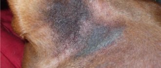
Redness and a pustular rash appear on the skin. A characteristic sign is thickening of the skin with changes in pigmentation - black spots. Due to damage to the sebaceous glands, a strong unpleasant odor appears.
The diagnosis is considered established after laboratory tests. The fingerprint smears are dried in air or fixed in the flame of an alcohol lamp and stained with a Gram stain. Microscopy reveals oval-shaped mushrooms. The diagnosis is confirmed by culture.
For local treatment, special shampoos Nizoral and Lactaderm are used. A chlorhexidine solution is applied to the damaged areas twice a day. To suppress fungi, ketoconazole is used systemically, daily for 3 weeks.
Preventing this fungal skin infection in dogs is quite simple. The infection develops with reduced resistance, so normal housing conditions will save the animal from problems.
What can you do at home?
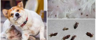
If you notice brown or black dots on your dog’s stomach, remember when and with what the extreme treatment against ectoparasites (fleas, ticks) was carried out.
Your actions:
- Carry out a comprehensive treatment of the premises and the animal.
- Deltsid anti-parasite concentrate (smells very strongly, requires long-term ventilation) or ParaStop , similar requirements apply to it (remove all animals, close windows tightly, treat surfaces, leave for exposure, ventilate). The duration of the protective effect of the latter is up to 6 months from eggs and larvae.
- For dogs, you can use tablets or drops. Anti-flea shampoos and collars are useless when infested. The most popular and effective flea drops: Advantix, Advocate, Advantage, BlochNet, Bolfo, Vectra 3D, PrakTik, Frontline . All treatments should be carried out in a therapeutic dosage - twice.
- Pills. At the moment, 3 brands of tablets are produced: Bravecto (valid for 12 weeks), Simparica and Nexgard (valid for 30 days). All drugs are absolutely safe and often help get rid of the problem better and faster. Bravecto, in addition to fighting fleas, is used in the treatment of demodicosis and protecting the animal from ticks.
- Visiting a veterinarian. Of course, a general veterinarian can provide advice, but it is better to consult a veterinary dermatologist.
To enlarge the picture, click on it
Comedones - damage to the sebaceous glands
When hair follicles are clogged with sebaceous gland secretions and dead epithelium, skin lesions are formed that look like black dots. This pathology is common in hairless dog breeds due to the high activity of the sebaceous glands. In other animals, comedones are less common and more often as a secondary disease.
A common cause of blackheads is hormonal imbalance. The thyroid gland normally regulates the formation of sebum and hair growth. Metabolic pathology and hormonal disorders lead to increased sebum production and the formation of comedones. There is a genetic predisposition to this disease in Schnauzer dogs - they develop rashes along the spine.
Black dots appear when dogs are affected by demodicosis - mites penetrate the skin and hair follicles, causing dermatitis.
Shampoos containing benzoyl peroxide are usually used for treatment. This product thoroughly cleanses the skin, removing formed comedones and preventing the appearance of new ones. Often the disease is complicated by a secondary infection, then systemic therapy is used - antibiotics, sulfonamides, skin treatments with antiseptics, anti-inflammatory drugs.
Caring for a dog during rehabilitation
Comprehensive care for your dog during the rehabilitation period includes:
- Once every three days, the dog is bathed using the above-mentioned keratolytic agents.
- The pet's fur is regularly combed, trimming it around the comedones.
- To prevent the animal from gnawing on damaged areas, a surgical collar is put on it.
- Three times a week, boiled sea fish, rich in polyunsaturated omega-3 fatty acids, is added to your pet’s diet. This has a beneficial effect on the speed of skin regeneration.
When is the risk of complications during self-care minimal?
You can try treating mastitis in your dog at home under the following general conditions:
- the gland is slightly swollen, there is no severe deformation;
- no increase in general body temperature;
- there is no severe pain that causes the dog to whine;
- milk can be expressed independently by hand, there are no signs of blockage of the milk ducts;
- no blood or purulent discharge;
- the inflammation did not cover the entire gland, but only its individual parts;
- You can notice improvements when using traditional medicine and home remedies during the first two days.
For the following symptoms (types) of mastitis in dogs, attempts to provide self-help are acceptable:
catarrhal mastitis:
| serous mastitis:
|
How to treat (if there is no veterinarian)
- Complete rest for a sick dog. If the individual has long hair, then it makes sense to cut off the hair around the nipple, exposing the affected part as much as possible for easy access and physiotherapeutic procedures.
- If there is no stagnation of milk, then the puppies are not weaned, applying more active and stronger ones specifically to the diseased lobes. If stagnation is detected, then the milk should be expressed by hand, and the puppies should be transferred to artificial feeding. The milk that stagnates inside literally turns sour and mixes with inflammatory secretions - not a single puppy, even the hungriest one, will suck such milk. Moreover, this can be fraught with digestive disorders in the offspring.
- Dairy products, meat and liquid soups should be completely excluded from the dog’s diet, and the amount of liquid consumed should be reduced (allow drinking only after meals). You can switch the animal to dry food during treatment.
- You should express milk only after a massage. Massage means stroking movements on the mammary gland, lightly pressing on it with your fingers so as not to cause pain to the dog. Particular attention should be paid to areas with compactions - dense areas of the gland that are clearly visible to the touch. You need to massage them until you feel them soften.
- Dogs' nipples have almost twice as many ducts as cats', so it's possible to express milk by hand (though not as easy). After massaging the entire gland, they smoothly move on to expressing, making stroking movements of the nipples from the base to the end, lightly pressing them with their fingers. In any case, it will be unpleasant for the dog, but you need to try not to cause pain (it is advisable to do everything with an assistant). There is no need to try to express all the milk from the breast; it is enough to do this until the milk lobe around the nipple collapses and becomes softer.
You can try to express milk with a homemade breast pump made from an ordinary disposable syringe (the volume of the syringe is selected according to the diameter of the nipple so that it passes freely and does not end up tightly). The piston is removed. The top of the syringe with the spout on which the needle is placed is cut off. The cut edges are aligned with fire to prevent skin injury and to ensure maximum adherence of the syringe to the skin. The nipple is inserted into the syringe, pressed tightly against the skin and the plunger is pulled - the milk will begin to be expressed a little at a time.
Before expressing, you can give your dog no-shpa at the rate of 40 mg (1 tablet) per 10 kg of weight. This will help relieve possible spasms of the milk ducts and make the procedure easier.
- If the local temperature is high (the gland is hot to the touch), cooling compresses are made - for example, you can apply a beaten and mashed chilled cabbage leaf or cool lotions from medicinal herbs: sage, chamomile, raspberry leaves, chamomile. Pour 200 ml of boiling water over 1 tbsp. any specified herb and leave for up to half an hour in a closed container. Then cool to the desired temperature, moisten gauze pads and apply to where it hurts. The top can be tied with a bandage (not too tight!). Change every time the napkin gets hot or every 2-3 hours.
- After the temperature has normalized, you can begin to make warm compresses from the same herbs as during cooling. The only difference will be in temperature - with warming compresses, the temperature of the solution should be 37-38°C. Important: it is forbidden to heat the mammary gland if there is even the slightest suspicion of purulent inflammation!
- You can use ichthyol, fir or camphor oil, which is gently rubbed into the surface of the skin of the diseased breast lobe, and then bandaged with a cellophane lining to obtain some greenhouse effect.
- Unlike cats, dogs can be given a thin iodine grid on the diseased mammary lobe - the distance between the “grids” is at least 1 cm, the frequency is once every 3-4 days. This is done with a cotton swab or a match with cotton wool wrapped around it. Lightly soak in iodine so that the stripes are slightly dark yellow and do not spread.
Further development of the disease
If you don't take your dog to the vet, things will get much worse:
- Small ulcers and boils burst, the chin area is sometimes completely covered with drying pus and ichor.
- As the animal’s immunity weakens, increasingly larger papules and boils appear.
- Bursted boils and papules do not heal; large ulcers develop in their place.
- Due to pain and intoxication, the animal partially or completely loses its appetite.
Diagnostic techniques
Everything is quite simple here. Since the symptoms are quite obvious, everything is usually limited to a simple external examination. The owner plays a significant role in this “action”. You must tell the specialist about the dog’s feeding conditions, changes or lack of appetite, diarrhea, vomiting, etc. If you have tried to treat your pet with something, do not be shy - if you tell the veterinarian about it in time, there will be much fewer problems later.
As a rule, specialists take a sample of pathological material in the form of a scraping and/or aspirate (pumping out the contents of the acne with a syringe)
. All this is used for further microscopic examination, or is sown on nutrient media for the purpose of growing a culture of the pathogen.
Malasseziosis
This name hides an interesting pathology of yeast origin. It is interesting because the causative agent of malassezia is not only not dangerous for an animal under normal conditions, but is also a common representative of the symbiotic microflora of the skin. But with a significant deterioration of immunity, yeast can cause illness. As a rule, such an outcome is either a consequence of a long, long and severe pathology of a different origin, or a long, unreasonably prolonged supply of corticosteroids, antifungals and antibiotics.
In addition, the first lesions of Malassezia never appear on the abdomen. They always occur between the toes and on their pads, less often in the genital area of the animal. And only subsequently do the lesions spread to the stomach, and sometimes to other parts of the pet’s body, including the face.
Symptoms of the disease are as follows::
- Since the first foci of the disease appear on the paws, you need to look at them. Loose, whitish-yellow growths appear between the fingers of Malassezia.
- A strong and rather specific odor emanates from the lesions. Some compare it to the aroma of old cheese, others think it is similar to the “amber” from equally old socks... In any case, it is difficult not to notice this sign.
- At first, the fungus does not cause any particular inconvenience to the dog, but soon pain and itching appears. The dog tries to lick these places, rubs them with his paws (which only worsens the condition of the skin and increases pain).
Treatment
When is self-help at home acceptable?
Treatment of mastitis at home can be done only in mild forms and non-purulent course. In all other cases, self-medication is unjustified and can have serious consequences. In any case, a specialist will advise you in detail on what to do if your dog has mastitis.
Action with puppies during mastitis treatment
Puppies can only be allowed to see a sick mother if they have serous and catarrhal forms. In these cases, they will help alleviate the condition of the nursing bitch. As soon as purulent, purulent-catarrhal or fibrinous forms are detected and pus or fibrin is detected in the milk, the puppies are weaned and transferred to artificial feeding. Contaminated milk can poison the offspring and lead to its death!
What happens if mastitis is not treated or treated incorrectly?
In case of prolonged (chronic) inflammation of the mammary gland and improper treatment (or refusal), the following consequences are possible:
- degeneration of glandular tissues with the formation of tumors (both benign and malignant);
- loss of secretory function and forced removal of the gland;
- death of the pet from general intoxication of the body;
- death of the offspring from poisoning with milk contaminated with bacteria.
Skin infections
Skin infections may occur on their own or may be secondary to an allergy. Regardless of the cause, if there has been an infection, it can lead to hyperpigmentation. Bacterial infections are the most common causes of skin infections.
- Mange (demodex, sarcoptic mange) can cause darkening. Demodex lesions are usually local.
- Sarcoptus usually becomes generalized. The discoloration may go away after scabies is treated, but it will remain after severe cases.
- Ringworm (dermatophytosis) can also cause temporary or permanent hyperpigmentation.
Veterinarians will ask the following questions:
- Why is the belly red or black?
- Where did the itching come from? How bad does it itch?
- Is there a foreign smell?
- Does it only itch at certain times of the year? (Allergy?)
The study is carried out by analyzing the medical history and diagnostics, for example, scraping, cytology, biopsy. If you adopt a dog that is healthy, not itchy anywhere, but has obvious dark areas, he probably suffered from illnesses such as allergies when he was little.