Acanthosis nigricans in dogs is a skin disease in which the animal experiences increased accumulation and production of melanin, baldness of the affected area where pigmentation is observed, and thickening of the epidermis. The violation is caused by several reasons. Treatment of a dermatological disease is carried out under the supervision of a veterinarian, and the factor that triggers acanthosis is necessarily eliminated.
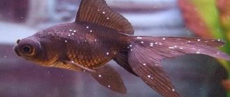
Typical location of acanthosis nigricans in the armpit
Characteristics of acanthosis nigricans
Acanthosis nigricans is a dermatological disease. Associated with dysfunction of the sebaceous glands. It manifests itself as a change in healthy skin to localized dark, rough folds, between which papillomas develop. The main places of damage: axillary area, neck, groin area, abdomen. Small spots and dots may appear in these places. If left untreated, the disease affects the entire body of the animal.
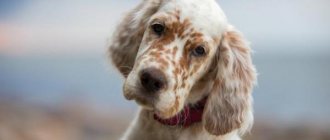
Acanthosis nigricans in a dachshund
General symptoms
Regardless of the type of disease, the clinical picture will be approximately the same. Initially, there is a barely noticeable darkening of the skin in the intergluteal region, on the abdomen, ears and inside the folds (head, limbs). Over time, the affected areas become more visible, standing out as black against the background of light skin.
Next, the skin changes density, becoming rough and hard to the touch. Growths of heterogeneous shapes appear in the folds. Sometimes this deformation is accompanied by a characteristic putrefactive odor. In parallel, alopecia may develop - gradual or, conversely, rapid hair loss in problem areas. The dog becomes restless, itches endlessly, does not allow darkened areas to be touched, and whines from pain and itching.
Causes of occurrence on the skin
The disease has three types, which determine the causes:
Primary
Affects dachshund dogs. The disease usually begins to develop at an early age.
Causes:
- hereditary factor;
- endocrinological diseases;
- kidney disease.
Secondary black
Develops predominantly in smooth-haired dog breeds.
Causes:
- neoplasms of various origins (benign and malignant);
- diseases of internal organs and systems;
- dysfunction of the genitourinary system;
- thyroid diseases;
- staphylococcal infection;
- chronic stress;
- overweight;
- poor environmental living conditions.
Pseudoacanthosis
Has similar symptoms. It is a skin rash (chronic or temporary), similar to skin changes like acanthosis.
Causes:
- allergy;
- dermatitis of various origins;
- hormonal imbalances;
- overweight.
In the localized area, the fur completely falls out, and an unpleasant odor begins to emanate from the pet.
What complications may there be?
Despite the fact that with timely treatment, acanthosis nigricans can be cured or its spread can be stopped, if the owner does not contact a veterinary clinic in a timely manner, the pet may develop a number of complications.
What could be:
- Formation of deep ulcers and abscesses. Occurs when the animal is restless, scratching spots and infection gets into the wounds.
- Baldness, roughening of the skin over large areas of the body.
- The appearance of other problems with the dermis, which aggravate the course of the underlying disease.
- Possible development of oncological processes in the dermis, both benign and malignant.
- Blood poisoning (septic phenomena) due to infection in the wounds. In this case, the lack of urgent veterinary care leads to death.
If the dog is constantly worried, the spots cause her discomfort, this affects her quality of life, the appearance of anorexia (refusal to feed), and weight loss. Complete apathy or, conversely, aggression cannot be ruled out.
Symptoms of the disease in a dog
The main symptom of the disease is damage to the skin.
The process occurs in several stages:
- At the initial stage, the skin darkens and becomes rough. Does not cause discomfort to the pet. Primary acanthosis ends at this stage.
- Next, the skin becomes rougher, forming a fairly hard crust, almost black in color. Rough skin is formed by folds, between which growths of different sizes form. The hair in this area becomes damaged and falls out due to constant brushing. Bald patches form.
- The next stage is infection of the damaged areas. This creates a characteristic unpleasant odor. The inflamed areas are painful and cause significant discomfort to the dog.
The second and third stages of the development of acanthosis nigricans are characteristic only of secondary acanthosis nigricans.
Another symptom at these stages is itching and the dog scratching the affected areas.
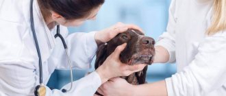
When acanthosis nigricans in dogs tends to progress, more aggressive therapy must be resorted to
Main symptoms
The development of pigmentation can be sluggish or, conversely, with rapid development, depending on the symptoms that cause it.
Signs of genetic pigmentation can include:
- single or groups of dark spots on the dog’s stomach, limbs, body, as well as on the eyelids and oral mucosa;
- pigmentation in the armpits and groin without signs of inflammation.
Signs of acquired pigmentation and ongoing inflammatory processes are:
- oddly shaped spots that change in size from small to noticeable changes in skin color;
- inflamed dark or red spots;
- the spots become thicker, itchy, and the skin becomes smoother;
- irritation due to friction in the armpits;
- depigmentation or pigmentation of the pet’s nose;
- Changes in coat color, for example, infectious pathogens cause light coats to turn rusty brown.
Diagnosis of small dark spots (dots)
All diagnostic research occurs in several stages:
Features of pathology and description
It is believed that the disease is based on genodermatosis, in which excessive hyperpigmentation occurs. But acanthosis of secondary origin is much more common, but its causes have not yet been established, although the study of this issue continues. The disease is considered a type of anti-inflammatory hyperpigmentation.
Causes of acanthosis nigricans may include:
- Inflammatory reactions in the most delicate groin and axillary areas.
- Endocrine diseases, incl. hypothyroidism, hypersecretion of corticosteroids.
- Obesity, when a dog’s weight is diagnosed above normal and as a critical body mass.
- Nutritional allergies due to improper feeding.
- Infections of the upper layers of the dermis.
Important! Contrary to popular belief among owners, acanthosis nigricans is not contagious. It is not transmitted from animal to animal, much less to humans. Another thing is that a dog with damage to the dermis does not look the best, so at the first signs of acanthosis you need to urgently contact a veterinary clinic.
Treatment of the disease
A complete cure for primary acanthosis is impossible.
Therapy methods are aimed at reducing the unpleasant manifestations of the disease to a minimum.
In this case, hormonal drugs and local remedies are prescribed.
If the dog is not bothered by the manifestations of the disease (there is no itching or inflammation), i.e. The problem is purely cosmetic; no therapy is prescribed.
In case of secondary maintenance therapy consists of:
Primary pathology, which is caused by genetic origin, cannot be completely cured
Breeds susceptible to pigmentation or coat color changes
No dog breed is immune from manifestations of pigmentation, especially in adulthood.
But there are breeds in which genetic abnormalities were monitored and their course was monitored - these are pugs (lentigines) and dachshunds (acantosis nigricans).
A tendency to lentigo has been noted in dwarf schnauzers and sharpeis.
German Shepherds are prone to acquired pigmentation. These are bacterial infections affecting the snout and lupus vulgaris.
Scottish Shepherds are predisposed to lupus vulgaris, which also affects the animal's face.
Chow chow and Akita Inu are predisposed to an acquired autoimmune disease that affects the skin with inflammatory purulent discharge.
Loss of pigmentation in the area of the nose and lips is caused by inflammation of the front part of the eye in breeds such as Akita Inu, Samoyeds and Siberian Huskies.
Doberman Pinschers, Rottweilers, Siberian Huskies, Alaskan Malamutes and Labrador Retrievers are prone to depigmentation (coloring the coat white).
Poodle, Yorkshire, Silky and Bedlington terriers, Old English Sheepdog are breeds that, due to a malfunction of the sebaceous glands or during the recovery period after an illness, may experience a change in coat color.
Consequences of the disease and complications
Complications and consequences of the disease occur in the absence of timely correct treatment or incorrect therapy.
Treatment must be prescribed by a veterinarian. Self-prescribing medications to a dog is unacceptable.
Illiterate use of pharmaceuticals can aggravate the situation, and a large number of side effects can worsen the pet’s health.
It is impossible to completely cure, but if you do not fight the symptoms, the likelihood of inflammatory foci occurring is 100%.
Injured, cracked skin is a favorable substrate for the development of bacteria and fungi. Which in turn leads to complex infectious diseases that affect the entire body of the animal. Further, if there is no treatment, the pet dies.
What to do at home
The appearance of spots on the skin, changes in coat color in dogs - all these insidious abnormalities cannot be treated on your own.
Without specific diagnostics in a veterinary clinic and finding out the main cause of the changes that appear, it is impossible to alleviate the animal’s condition.
Therefore, at home you should follow all doctor’s prescriptions, and if the diagnosis of lupus is confirmed, you should protect your dog from exposure to ultraviolet rays.
The use of ointments and medications selected by a doctor will speed up your pet’s recovery.
Caring for a sick dog
Features of caring for a sick dog are related to alleviating the pet’s condition and maintaining sanitary conditions in the animal’s place of residence.
It is necessary to carry out all medical procedures strictly as prescribed by the veterinarian: treat damaged skin, wash the animal.
Carefully ensure that the dog does not scratch the affected areas (give sedatives and antihistamines in a timely manner).
Maintaining cleanliness in the area where the dog lives is necessary to prevent infection of the injured skin.
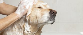
Antimicrobial shampoo will help with inflammation
Forms
Pathology has 2 forms:
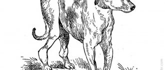
- Primary . Another name for it is genodermatosis (a skin disease that is genetically determined). A huge number of dog breeds suffer from this disease, but dachshunds are most susceptible to it. They develop all the characteristic signs of acanthosis nigricans before they are one year old.
- Secondary . It is called post-inflammatory hyperpigmentation. This disease occurs in almost all breeds; age in this case does not matter. Post-inflammatory hyperpigmentation occurs against the background of diseases that are accompanied by inflammatory processes located in the groin or axillary region, obesity, and endocrinopathy.
Many people believe that acanthosis nigricans is contagious. However, in fact, they are deeply mistaken, since this disease is genetically determined, that is, specific: it cannot pass from one sick dog to another healthy one.
Clinical signs of acanthosis nigricans
Symptoms of the disease begin to appear in puppies one year of age, regardless of the type of occurrence.
As the disease progresses, the hair begins to fall out.
A symmetrical change in pigmentation is visible under the armpits of the limbs. Ignoring the symptoms leads to thickening of the skin in the affected areas, hair loss and the appearance of signs of lichen. Further progression of the disease is characterized by similar symptoms on the back of the paws, under the neck, and inside the ears. Lack of treatment provokes infection in the diseased area and symptoms of secondary seborrhea occur, severe itching appears.
Diagnostics
Diagnosis consists of differentiation from similar diseases, which include:
- food allergies;
- contact dermatitis;
- atopy;
- hypothyroidism;
- hyperadrenocorticism;
- hyperestrogenosis.
Confirmation of the diagnosis is carried out by biopsy of skin pieces . If acanthosis is not treated, the disease becomes chronic and manifests itself as seborrhea. Scaly growths of various shapes and structures appear on the surface of the skin. Itching may be present. The dog's fur becomes dull and gives off an unpleasant odor. Over time, the scales turn into plaques and the skin turns black.
If acanthosis is not treated, the disease will become chronic.
Disease progression
The progression of the disease is expressed in extensive damage to an increasingly larger part of the body.
- The dog itches all the time , sometimes scratching the skin until bleeding wounds form.
- The dog becomes nervous , does not give in to hands, when trying to touch it growls and tries to bite, which is explained by the pain of the formations.
- The appearance of a bright red border along the edges of the lesions indicates that infectious flora is developing in the lesions.
During illness, the dog is constantly itching.
Lentigo
Black spots on the skin may be a cosmetic defect such as lentigines. There is no effective therapy for this form of pathology. Lentigo can be localized in the dog’s abdomen or limbs. Less commonly, a cosmetic defect is diagnosed in other areas of the animal’s body.
Most often, lentigo appears in middle-aged and elderly dogs.
The small dark spots that appear begin to gradually increase in size over several months. Not only does the size of the spots grow, but their number also increases. After a certain period (for each animal individually), the growth of spots stops.
From the outside, lentigines look like flat, well-defined spots that do not protrude above the surface of the skin. True lentigo in a dog does not itch, does not provoke itching, and the surface of the spot is smooth. If the black spot has a lumpy structure or thickenings are present, it is recommended to immediately contact a veterinary clinic for advice and an accurate diagnosis.
Pathogenesis
Acanthosis nigricans in humans is characterized by symmetrical hyperpigmented foci of hyperkeratosis and papillomatosis , predominantly affecting skin folds. Initially occurring hyperpigmentation against the background of dryness and roughening of the skin subsequently acquires a grayish-black slate tint, folding of the skin, numerous and small villous and warty formations are formed. The spread is usually gradual and symmetrical over the years, and may be accompanied by dry hair, nail dystrophy, and increased keratinization of the surfaces of the palms and soles. The changes are quite often caused by the long-term effect of insulin on keratinocytes - the binding of insulin molecules on the surface of the epidermis to receptors for insulin-like growth factor.
At its core, malignant acanthosis nigricans is an obligate paraneoplastic dermatosis . Paraneoplastic processes in the body cause skin lesions and a complex of symptoms. The pathogenesis is based on the processes of cellular proliferation of keratinocytes caused by blastoma growth. Chronic inflammation also creates a favorable background for the development of neoplasia. In this case, the inflammatory response is usually directed not only at the tumor, but also at molecules that are expressed in the epithelium and cause paraneoplastic changes.
Prevention
As for prevention, genetic acanthosis cannot be prevented.
- If a dog has a genetic predisposition to this disease, then the disease cannot be stopped, only progression and complications can be prevented.
- Acquired pathology can be avoided if your pet is vaccinated in a timely manner and the occurrence of provoking factors is prevented.
- Monitor the sanitation of the habitat to prevent possible infection.
Timely vaccination will prevent the occurrence of the disease.
Diseases caused by pathogenic fungi
Dermatophytoses
Skin diseases resulting from parasitism by pathogenic fungi of the genera Microsporum and Tpichophiton are collectively called dermatophytoses (dermatomycosis). Dermatophytes have the ability to infect healthy hair or skin. The infected part of the hair is very fragile and often breaks off, so the hair in the affected area has the appearance of being cut off (hence the outdated name “ringworm”), up to complete baldness. In addition to the hair shaft, areas of interfollicular keratin can also be affected, which is manifested by peeling of the skin surface. The severity of inflammation varies. Infection with non-host-specific fungal species causes a stronger inflammatory response than with host-adapted species. The hyphae of some (not all) Microsporum species produce a substance called pteridine, which can absorb ultraviolet rays and fluoresce. This property underlies the luminescent diagnosis of microsporia. However, the absence of fluorescence does not exclude dermatophytosis.
It is believed that the cause of dermatophytosis in cats in 95% of cases is Microsporum canis. The remaining 5% of cases are caused mainly by Microsporum gypseum and Trichophiton mentagrophites. Microsporum canis is well adapted to cats as hosts, so the disease is often asymptomatic. It is very difficult to isolate the pathogen from healthy animals that have not previously suffered from microsporia, while the carriage level in cats suffering from dermatophytosis reaches 100%.
Dermatophytosis is more common in kittens and occurs in a more severe form. In adult cats, the disease is more common in individuals with weakened immune systems, pregnant or lactating. Sick cats pose a danger to humans and other animals.
Dermatophytosis is much less common in dogs than in cats. In most cases, Trichophiton mentagrophites, Microsporum canis, Microsporum gypseum are responsible for the occurrence of dermatophytosis in animals of this species. Clinical signs of dermatophytosis are very diverse and are not always limited to the classic picture of the disease. In most animals, the most typical manifestations are erythema, alopecia, scaling and crusting. Lesions are localized on the scalp and neck, less often at the base of the tail and limbs. The spots are small at first, gradually expand and reach a significant size. Hair loses normal pigmentation, becomes brittle and is easily epilated. The skin in the affected area is dense, red-brown or grayish in color. Animals scratch the affected areas, which often contributes to the transfer of the disease from the head to the limbs. As a rule, the itching is mild. The exception is infection caused by Trichophiton mentagrophites. Dermatophytosis can occur in conjunction with other diseases, mainly ectoparasitic: demodicosis (in dogs) and notoedrosis (in cats).
Malassezia dermatitis in dogs
Recently, a number of skin diseases in dogs (atopic dermatoses, otitis externa) have been complicated by yeast fungi of the genus Malassezia, especially Malassezia pachydermatis. Dogs of all breeds are susceptible to Malassezia dermatitis, but basset hounds are particularly susceptible. Skin lesions that are associated with Malassezia pachydermatis may be localized or generalized. Affected areas usually include the external auditory canal, muzzle, ventral neck, axillary cavities, groin area, and interdigital skin folds. Most often, the disease is characterized by erythema, alopecia, and dry or oily seborrhea. In chronic cases, lichenification and hyperpigmentation are observed. The itching varies from mild to extremely severe. Skin lesions are often accompanied by an unpleasant odor, especially in places such as the neck, axillary fossae, and ears.
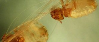
Acarodermatoses
Tick-borne dermatoses of dogs and cats occupy a significant place among skin diseases in these types of domestic animals. Diseases caused by these arthropods are usually accompanied by severe itching, scratching, baldness, and secondary pyoderma, which not only causes suffering to the sick animal, but also greatly upsets its owners. In addition, sick dogs and cats are a source of disease for other animals and for people. Therefore, the closest attention must be paid to the treatment of these dermatoses. For veterinary dermatological practice, acarodermatoses such as notoedrosis , sarcoptic mange , otodectosis and demodicosis . These diseases affect both dogs and cats, but with varying frequencies. Thus, notoedrosis and otodectosis are more often recorded in cats, sarcoptic mange and demodicosis - in dogs.
Atopic dermatitis
Atopy is a hereditary predisposition to the formation of antibodies against allergens from the environment (pollen, poplar fluff, house dust, etc.). Since atopy is a polyetiological disease with a variety of clinical manifestations, its diagnosis and treatment pose a certain difficulty for veterinarians. In terms of frequency of occurrence among all allergies, atopic dermatitis is second only to allergic dermatitis from flea bites. Often the latter accompanies the former, complicating and confusing the clinical picture of the disease. Food allergy, and in dogs also pyoderma, can also contribute to the complication of the disease. The clinical manifestations and diagnosis of atopic dermatitis in dogs and cats are largely similar, but there are also differences.
In dogs, atopic dermatitis most often occurs between the ages of 1 and 3 years and affects from 3 to 15% of the entire population of animals of this species, regardless of gender. Breeds susceptible to this disease include terriers (WHWT, Scotch, Fox), golden and Labrador retrievers, boxer, cocker spaniels, German shepherd, Shar-Pei, Dalmatian, English bulldog, miniature schnauzer, Irish and English setters. The most typical clinical signs of atopic dermatitis in dogs are pruritus, alopecia, erythema, hyperpigmentation, and lichenification, which are found on the face, feet, chest, ears, abdomen, and tail. Depending on the source of the allergen, atopy can be seasonal or cause trouble for the animal and its owner for most of the year.
Dogs with atopic dermatitis are more likely than others to be affected by a yeast infection (Malassezia), which is facilitated by inflammation and oily seborrhea. In the interdigital spaces, ideal conditions for the proliferation of fungi are created due to increased humidity and relatively higher skin temperature in these areas.
Changes in skin pigmentation
Pigment spots can be of any color (from dark black to reddish). Each type is caused by a specific condition or disease.
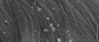
Black dots
The appearance of black spots on a dog's skin is most often caused by lentigo. It consists of single spots or group spots of dark black color. Their occurrence is caused by macular melanosis, that is, excessive accumulation of melanin in the body. With canine lentigo, the appearance of flat, hyperpigmented spots of a benign nature is observed. Their localization is the groin area or abdomen.
Determining the causes of black spots
Determining the etiological factor of the disease and correct diagnosis are necessary for the rational organization of treatment. It is necessary to completely collect anamnesis, conduct a clinical examination and prescribe functional diagnostic methods. A big mistake is prescribing symptomatic treatment. In addition, the symptomatic picture is often blurred by the use of antibiotics and other medications. First of all, you should familiarize yourself with the complaint of the owner who has noticed changes in the behavior and appearance of the animal. At the same time, check the general condition of the pet:
- behavior (activity, reaction to stimuli) – apathy is characteristic of hypothyroidism;
- changes in appetite, increased thirst - occurs with improper use of steroid medications, hyperadrenocorticism;
- the presence of signs of digestive disorders occurs during intoxication;
- nervous clinic is also characteristic of hyperadrenocorticism;
- conjunctivitis and other eye diseases due to allergies;
- otitis, diseases of the ear canals due to allergic reactions, metabolic pathologies;
- exhaustion as a consequence of neoplasms, metabolic disorders, chronic diseases.
It is important to find out the nature of your pet's environment. Are there other animals in the house, or can the dog come into contact with stray animals? It is also worth finding out the presence of similar signs in people - fungal diseases and mites are common. It is necessary to clarify the place where the pet is kept (bedding, room).
Then they begin to study the location of the pathological process. It is necessary to clarify the location of the spots and the primary source of their appearance. Perhaps the spots have a seasonal tendency to appear (allergies, flea dermatitis) or the disease progresses. The presence of itching is determined and whether the spots cause anxiety to the dog. It is important to know the previous treatment - the use of special medications, flea and tick treatments .
A skin examination includes examination of all integuments, and not just in areas with pronounced lesions. Mucous membranes are also studied. The nature of the coat is noted - oily skin, hair color and structure, and the presence of bald patches. The skin on the abdomen, neck, perineum, and base of the ears is carefully examined. During the examination, the temperature of the skin, the condition of the surface, and the presence of various types of damage are noted.
Taking an anamnesis and a complete clinical examination allows you to make a preliminary diagnosis and choose a direction for further examination of the dog. A wet paper test should be performed to check for fleas.
If necessary, skin scraping is done in the area of the pathological process. It is carried out with a scalpel at the border of the affected and healthy areas. Pathological material is decolorized with an alkaline solution and examined under a microscope. Cytological examination material is also collected for bacterial and fungal infections.