- October 8, 2018
- Ophthalmology
- Evdokimova Irina
The state of the organ of vision can tell a lot about the health of pets. Cloudiness of the cornea in a dog indicates a serious problem in the body. This symptom can be observed in a variety of pathologies that require immediate treatment. In advanced cases, damage to the cornea can lead to blindness. In this article we will consider in detail the possible causes of cloudy eyes and methods of treating pathologies.
Non-pathological causes
Corneal clouding in dogs is often observed in old age. In old age, the animal's eyes may be covered with a whitish film. This is due to age-related changes in the corneal layer. A sign of aging is also the appearance of a white spot near the pupil. This is not a sign of pathology and does not require treatment.
In blue-eyed dog breeds (such as huskies), the cornea may fade with age. In old age, these animals' eyes become almost white. This is normal and does not indicate ophthalmic diseases.
Symptoms
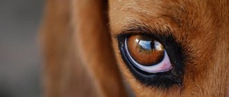
At the first signs of pathology, the dog begins to squint. After some time, the surface of the eye becomes cloudy, acquires a whitish or slightly blue tint, and becomes matte.
With any pathology of the lens, the cornea remains transparent, and a cloudy spot can be observed in the area of the pupil.
After injury, turbidity is observed in the anterior chamber of the apple. This is a consequence of the accumulation of pus or blood discharge.
Possible diseases
However, cloudy eyes in dogs are not always caused by age-related changes. This symptom can also occur in young animals, including puppies. Most often this is associated with the following pathologies:
- glaucoma;
- cataracts;
- inflammatory eye diseases (keratitis, uveitis, conjunctivitis);
- consequences of injuries;
- dystrophic and degenerative changes in the organ of vision;
- piroplasmosis.
Next, we will consider in detail the symptoms of the above pathologies, as well as methods of their diagnosis and treatment.
Glaucoma
Glaucoma is one of the most serious eye diseases in dogs. This pathology is characterized by increased intraocular pressure. Because of this, the animal's eyes look bulging. The cornea is covered with a cloudy film of grayish-bluish color. Glaucoma is often accompanied by severe pain. The pet becomes restless, whines and squints, especially in bright light.
Without treatment, glaucoma can lead to complete loss of vision. If an increase in intraocular pressure is accompanied by inflammation of the choroid (uveitis) or changes in the lens, then there is a danger of the appearance of malignant tumors in the organ of vision.
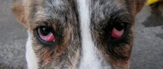
Cherry eye
“Cherry eye” - “cherry eye” - this pathology is often called “inflammation of the third eyelid”, “adenoma of the third eyelid”.
Dogs have three eyelids: two that are clearly visible and one extra eyelid (called the third eyelid), which is usually hidden under the inner corner of the eye. The third eyelid contains the gland that produces tears. Usually this gland is barely noticeable, but some dogs have a congenital weakness of the ligaments that hold it in place. When these ligaments fail, the gland jumps out of its normal position and looks like a “cherry” stuck in the inner corner of the eye. Because the condition often has a genetic basis, both eyes are usually affected over time. To treat cherry eye, the veterinarian performs simple surgery to return the gland to its normal position.
Cataract
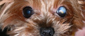
Cloudy eyes in a dog can be a sign of cataracts. This disease is more often observed in animals older than 6 years. With age, the tissues of the eye undergo sclerotic changes. Calcium salts are deposited in them. Because of this, clouding of the lens occurs.
At the initial stage, this disease can occur without any external manifestations. Then a blue or white spot appears on the dog’s pupil. As the pathology progresses, it increases in size. In advanced cases, the spot completely covers the pupil. This is accompanied by severe visual impairment. A sick dog moves very carefully and constantly bumps into objects.
In 80% of cases, cataracts are hereditary. A predisposition of some breeds to this disease has also been revealed. Poodles, terriers, miniature schnauzers, and golden retrievers most often suffer from clouding of the lens.
Animals with diabetes mellitus, autoimmune pathologies, and chronic infections often suffer from cataracts. This disease often occurs after an eye injury in a dog. Cloudiness of the lens can be a consequence of bruises, chemical and sunburns of the organ of vision.
Conjunctivitis
This disease is an inflammation of the mucous membrane on the inner surface of the eyelids. It occurs due to infection or dirt and dust particles getting into the eyes. An allergic form of conjunctivitis also occurs.
With this disease, there is severe redness of the whites of the eyes and increased tearing. The pet is constantly bothered by itching, which is why he tries to reach his eyelids with his paws.
Cloudiness of a dog's eye with conjunctivitis occurs due to the formation and accumulation of exudate. On the cornea and sclera you can notice a film consisting of mucous or purulent discharge. The edges of the eyelids become covered with a yellowish crust.
Keratitis
This disease is an inflammation of the cornea. It often occurs as a complication of conjunctivitis. The inflammatory process can move from the mucous membrane to the cornea in just a few days. Another cause of keratitis is insufficient functioning of the lacrimal glands. As a result, impurities and dust are not washed out of the animal’s eyes.
Eye injuries in dogs can also cause inflammation of the cornea. Through small scratches and abrasions, infection penetrates into the tissue. In this case, the inflammatory process is accompanied by suppuration.
With keratitis, spots form on the cornea. They look like pearly gray dots. A sick dog's eyes become extremely sensitive to light, causing the pet to constantly squint. The animal blinks frequently and appears restless. There is an increased secretion of tears and sometimes pus.
In advanced cases, ulcers appear on the cornea. Due to ingrowth of blood vessels, the sclera turns red. Such signs indicate a severe stage of the disease; without treatment, this pathology leads to loss of vision.
Keratitis most often affects small breeds of dogs: Pekingese, French bulldogs, pugs. This is due to the peculiarities of the anatomical structure of the head and eyes.
Types and course of the disease.
Catarrhal superficial keratitis in dogs
- the mildest form of superficial inflammation. Occurs after mechanical damage. Only the upper layer of the cornea is involved in the process. Cloudiness of the cornea occurs within a few hours, and sometimes it can disappear in one laziness. The surface of the cornea becomes rough and gray-bluish in color. Manifested by severe photophobia and lacrimation.
Purulent superficial keratitis
is a purulent inflammation of the upper layers of the cornea's own substance. Develops from catarrhal keratitis, not previously treated. It is also accompanied by photophobia, only more pronounced, pain and purulent discharge. The surface of the cornea becomes rough, swollen and whitish-bluish in color. Over a long period of time, vessels grow into the cornea. If this disease is neglected, superficial keratitis will develop into deep purulent keratitis or a corneal ulcer will develop.
Parenchymal keratitis
provoked by a bacterial infection, usually develops against the background of canine distemper, toxoplasmosis, can also be caused by trauma to the visual organ, often occurs in parallel with conjunctivitis and iritis (inflammation of the vessels of the cornea of the eye). This type is characterized by severe lacrimation, swelling of the cornea with the formation of spots and spots characteristic of this form.
Phlyctenulous keratitis
Caused by chemical poisoning or allergies. Most often found in collies and shepherd dogs. Large grayish-white bubbles appear on the cornea. Without veterinary care, they eventually burst, causing the cornea to turn reddish-gray. The third eyelid thickens and acquires a bumpy texture.
Ulcerative keratitis in dogs
is an inflammatory process in which the tissue of the cornea dies and defects (ulcers) form on it. In dogs, ulcers can be purulent, perforated or creeping. Ulcerative keratitis occurs as a consequence of other inflammatory processes of the cornea described above. Often develops due to penetrating wounds of the eyeball. Often the cause of this type of disease is a herpesvirus or bacterial infection. Treatment for ulcerative keratitis is quite long. But it must be timely.
Punctate keratitis
It occurs in dogs less often than other forms; the reason why it occurs is still unknown. Pearlescent spots form on the mucous membrane of the affected eye, but the dog’s vision and general well-being do not deteriorate.
Neurogenic keratitis
develops due to damage to trophic nervous tissue. Then a flat ulcer forms in the center of the cornea. The process is long and sluggish. Sick dogs may not notice discomfort for a long time. If the prognosis is favorable, the ulcer will heal, leaving behind a slight clouding. The addition of a secondary infection causes tissue perforation (formation of a through defect) and its destruction.
Uveitis
With this disease, the choroid of the eye (uveal tract) becomes inflamed. This structure is located between the cornea and the lens. Uveitis never occurs as a primary pathology. Inflammation of the eye vessels becomes a consequence of infectious diseases: leptospirosis, leishmaniasis, borreliosis, herpes. It is often observed in dogs infected with worms.
Uveitis is characterized by severe pain. The animal often whines and tries to touch its eyes with its paws. In some cases, strabismus is observed. The sclera of the eyes appears swollen and red. A large amount of opaque exudate is formed. You may notice clouding of the dog's cornea due to the accumulation of secretions.
This is a rather dangerous pathology. It is often complicated by the development of cataracts and glaucoma, as well as retinal detachment.
Belmo
Pet owners often confuse cataracts with cataracts. The external manifestations of these pathologies are similar: a white spot appears on the dog’s cornea. However, these diseases have different origins.
Cataracts are formed due to the deposition of calcium salts in the lens. A cataract occurs as a result of injury to the dog's cornea. Burns most often lead to this complication. The healing process is accompanied by the appearance of scars. A white spot remains at the site of damage. It does not increase in size. Over time, this formation turns yellow due to fat deposition.
What should you do if your dog’s eye becomes cloudy after an injury and a thorn appears? If the spot has formed on the periphery of the cornea, then it has virtually no effect on visual acuity. If a cataract appears in the pupil area, this can lead to complete or partial blindness. Conservative treatment is possible only at an early stage. In advanced cases, the animal requires eye surgery.
Types of lens opacities
To find out how a person with cataracts sees the world, imagine looking into the distance through a running stream of water. It’s as if you are standing on one side of a waterfall, and what interests you is on the other side. It is unlikely that under these conditions you will be able to view objects with a high degree of clarity. So the eye with cataracts perceives surrounding objects as if through a thick film.
Clouding of the lens gradually causes a change in the pupil of the eye, in which it becomes whitish rather than black. Such transformations are irreversible, but it is still possible to stop the progression of destructive processes.
There are several types of cataracts:
- Age-related - develops in adulthood, mainly after 50 years.
- Congenital type - a child is born with this form of disorder, and manifestations of the disease occur in the early stages of life.
- Traumatic - pathology arises after blows, bruises, mechanical and chemical damage.
- Radiation - this form of cataract is extremely rare and is caused by exposure to background radiation.
Dystrophic and degenerative changes
Corneal dystrophy is a hereditary disease. With this pathology, the division of epithelial and endothelial cells is disrupted in the animal. Sagging and a cloudy blue film appear on the cornea. Dystrophic changes are not accompanied by painful sensations, the disease progresses very slowly.
As an animal ages, salts and cholesterol are deposited in the cornea. This leads to tissue degeneration. Swelling and a gray-blue spot forms. First, it is localized in the lateral part, and then spreads to the entire cornea. At the initial stage, the pet does not experience any unpleasant sensations. But in the future, degeneration is complicated by ulcerative keratitis, which is accompanied by severe pain.
Piroplasmosis
Clouding of the cornea in dogs with piroplasmosis appears in the first days of the disease. The causative agent of this pathology is the simplest microorganisms - Babesia. They enter the dog's bloodstream when bitten by ixodid ticks. Babesia causes massive destruction of red blood cells, enlargement of the liver and spleen.
With piroplasmosis, the dog's eyes acquire a yellowish tint. The mucous membranes of the mouth are painted the same color. Such symptoms are a sign of hemolytic jaundice, which develops against the background of the death of red blood cells and an increase in bilirubin levels.
Piroplasmosis is also accompanied by the following manifestations:
- a sharp increase in temperature (up to +41 degrees and above);
- severe weakness;
- staining urine red or brown;
- vomiting;
- difficulty breathing.
This parasitic pathology is extremely dangerous. Without treatment, it can lead to the death of the pet from intoxication in the first days of the disease.
Diagnostics
If a dog's cornea becomes cloudy, it is necessary to take the animal to a veterinary clinic as soon as possible. Only a specialist can determine the exact cause of such a symptom. Animals are prescribed the following types of examination:
- examination of the organ of vision using an ophthalmoscope;
- measurement of intraocular pressure;
- swab from the eye for bacterial culture.
If piroplasmosis is suspected, it is necessary to take a blood smear for Babesia. In sick dogs, the causative agent of the disease is found in red blood cells.
Dry eye
When dogs develop a condition called keratoconjunctivitis sicca or dry eye, their tear glands produce fewer tears than normal. Tears perform important functions:
- removing potentially damaging material from the surface of the eye;
- nutrition of corneal tissue.
Mild cases of dry eye can be treated with an artificial tear solution, but medications that stimulate tear production (such as cyclosporine) are usually needed.
Drug therapy
So, we found out that there are many diseases that cause clouding of the cornea in dogs. Treatment will depend on the cause of the symptom. Drug therapy is effective for inflammatory diseases of the organ of vision. Let us consider in more detail the methods of treating such pathologies:
- Conjunctivitis. For this disease, antibacterial eye ointments and drops are prescribed. Before using them, the inner surface of the eyelids is washed with chamomile decoction. For severe itching, topical corticosteroids are indicated. If inflammation of the mucous membrane is associated with an allergic reaction, then antihistamines (Suprastin, Cetrin) are prescribed in tablets.
- Keratitis. In case of inflammation of the cornea, rinsing the eye with antiseptics (a solution of furatsilin or boric acid) is indicated. It is also recommended to instill drops based on sodium sulfacyl. To relieve inflammation, steroid hormones are administered by injection.
- Uveitis. For inflammation of the blood vessels in the eye, animals are prescribed corticosteroid ointments. These drugs penetrate the cornea and stop the inflammatory process. In severe cases, hormone injections into the conjunctiva are indicated.
Glaucoma can be treated conservatively only in the early stages. Animals are prescribed a course of diuretics, which are administered intravenously. Removing excess fluid from the body helps reduce intraocular pressure.
Treatment of corneal ulcers depends on their etiology. If erosion is a complication of bacterial diseases, then eye ointments based on erythromycin and tetracycline are prescribed. For viral infections, Idoxuridine and Trifluridine drops are used.
Non-surgical treatment of cloudy eyes in a dog due to burns and other injuries is possible only in the early stages of the disease. Vitreous injections will help get rid of the cataract. This medicine promotes the resorption of scars.
For piroplasmosis, dogs need a course of injections of the antiprotozoal drug Azidin-vet. This is one of the few products that can destroy Babesia. Pet owners in reviews note the effectiveness and safety of this medicine. It causes virtually no side effects. The yellow film on the eyes disappears after complete recovery and normalization of red blood cell levels.
How to save your eyesight?
To make a diagnosis, a veterinarian ophthalmologist carefully examines your dog's cloudy eyes using several techniques. These include:
- checking for strabismus;
- assessment of visual function and condition of the cornea;
- checking the blink reflex.
When visual function deteriorates, the animal bumps into objects or moves very carefully, avoiding possible collisions. The doctor assesses the amount of discharge and the size of the film formed. The condition of the lens is assessed using an ophthalmoscope and a slit lamp. Additionally, ultrasound, examination of the anterior chamber of the eyeball (gonioscopy) and measurement of intraocular pressure (tonometry) are prescribed.
In addition to standard procedures, it is possible to perform a Schirmer test and take a Seidel test with fluorescein. The first technique examines the amount of tear fluid. Filter paper is placed behind the lower eyelid of a four-legged patient. If it gets wet within 5 minutes, then the patient is healthy. If the piece of paper remains dry, the presence of pathology is confirmed.
The second technique is used to detect damage to the cornea. During the examination, the doctor applies a fluorescent solution to it and then presses it with a cotton swab. All actions are carried out under local anesthesia. If dark-colored leaks appear, a perforated ulcer is confirmed, that is, a lesion with a through hole. The dog urgently undergoes microsurgical sealing of the wound.
Depending on the diagnosis, treatment can be medicinal or surgical. In the first case, a dog’s cloudy eye is treated exclusively with medications, in the second, it is necessary to resort to surgery.
Drug treatment
Drug therapy is prescribed for the following pathologies:
- Conjunctivitis
. The patient is prescribed antibiotics. Treatment is carried out at home. For recovery, it is necessary to do regular rinsing with an antiseptic. After cleansing, eye drops are instilled, and after 15 minutes an antibacterial ointment is placed under the eyelids. The frequency and duration of procedures is determined by the veterinarian. If the reason lies in allergies, then, in addition to general recommendations, it is necessary to limit exposure to the allergen and take a course of antihistamines.
- Keratitis
. The lacrimal sacs are cleaned of accumulating pus with antiseptics. Depending on the form, keratitis is treated with eye drops, intramuscular injections of antibiotics, corticosteroids, antivirals or antihistamines. To prevent scratching, a special collar is put on the pet. On average, treatment takes 1.5 months. For advanced forms of keratitis, tissue therapy or superficial keratectomy (laser vision correction) is recommended.
- Uveitis
. Cloudy eyes in dogs caused by uveitis are treated with antibiotics, anti-inflammatory drugs and drops. All these drugs are used as auxiliary drugs. The main therapy depends on the underlying cause of uveitis. If the doctor diagnoses a high probability of developing glaucoma, then removal of the diseased organ is recommended.
- Belmo
. Most often, the thorn is eliminated according to the general scheme. Additionally, vitreous injections are used to promote resorption of the stain. In rare cases, they resort to surgical removal of the film or corneal implantation.
For more severe pathologies, drugs are used exclusively to eliminate symptoms.
Surgery
If drug therapy fails, the only option left is surgical intervention. At best, it restores the loss of visual function, at worst, it slows it down. Pathologies that require mandatory surgery include:
- Cataract
. A four-legged patient has a clouded lens removed using ultrasound. This method is called phacoemulsification. Ultrasonic waves transform the biological lens into an emulsion. After this, the doctor makes a microscopic puncture and removes the fluid. An implant is installed in place of the removed lens. The disadvantage of this therapy is the high price and the small number of qualified doctors. In the early stages, the development of the disease is slowed down by anti-inflammatory and antioxidant drugs. Treatment should begin immediately after the loss of the usual shine.
- Glaucoma
. Restoring original vision is impossible, so all actions are aimed at preserving the remaining vision. First of all, doctors lower intraocular pressure and perform cyclocryotherapy. This method involves using cold to reduce tear production. Additionally, medications are prescribed that normalize or inhibit the production of intraocular fluid: beta blockers, osmotic diuretics, carbonic anhydrase inhibitors, miotics. Most often, four-legged patients are admitted to the clinic with an advanced form of the disease. In this case, nucleation or evisceration is used. Both operations involve the removal of a painful organ, but with evisceration subsequent prosthetics are provided.
- Dystrophy
or
corneal degeneration
. Corneal dystrophy is eliminated by keratoplasty, which involves removing the affected area. Its disadvantage is the appearance of scars that impair visibility. A more effective method is a cornea transplant. But even in this case, a bluish film often remains. Also, the disadvantages of this operation include its price. Not all owners are able to cover the necessary expenses. If the disease is detected at an early stage, conservative therapy is possible. The animal is prescribed antibiotics, ointments and eye drops. Unfortunately, such treatment rarely produces positive results.
Elimination of erosion or ulcer depends on the form of the pathology. Typically, visual function returns after taking medications that dilate the pupil, non-steroidal anti-inflammatory drugs, antibiotics and drugs with an anti-collagenolytic effect.
In the chronic form, it is recommended to use surgical techniques:
- keratotomy - leveling the cornea with radial incisions;
- treatment with diamond bur – removal of damaged epithelium;
- keratectomy – removal of all damaged layers;
The best results are achieved by keratectomy. It is easier for renewed tissues to take root on a healthier surface. The disadvantage of this operation is the need for general anesthesia. For older animals and pets with weak hearts, only local anesthesia is recommended.
Operation
For cataracts and in the later stages of glaucoma, dogs are indicated for surgical treatment. These diseases respond poorly to conservative therapy. The following types of surgical interventions are performed:
- Lens replacement. This operation is performed for cataracts. The affected lens is removed from the animal and replaced with an artificial one. In this case, the eye tissue has to be cut.
- Phacoemulsification. This is a less traumatic method of cataract surgery. All manipulations are carried out through a small puncture. The affected lens tissue is sucked out and a rolled-up artificial lens is inserted in its place.
- Removal of the eye. This operation is performed in severe cases of glaucoma, if there is a risk of malignant tumors.
Dog owners respond positively to the treatment of cataracts using phacoemulsification. Animals tolerate this operation well. After replacing the clouded lens, pets regain lost vision. However, veterinarians emphasize that the question of the need for such intervention can only be resolved on the basis of a comprehensive examination. If the animal is diagnosed with infections, heart disease or retinal damage, then the lens cannot be replaced.
In case of degenerative and dystrophic processes in the cornea, as well as in the later stages of the cataract, surgical correction is performed. The affected tissue is removed surgically. However, after such an intervention, scars remain, which negatively affects the quality of vision. A radical treatment method is a corneal transplant. The changed tissues are removed from the dog and replaced with donor biomaterial or an artificial graft. This operation is very expensive and not available to all animal owners.
Treatment
Treatment of the disease very much depends on its cause and the moment of contacting a doctor. In ophthalmological problems in dogs, the symptom of which is cloudy eyes, one of the most important aspects of successful treatment is early detection of the pathology. If the diagnosis is made at an early stage, most problems can be solved with therapeutic treatment - drops and ointments. In advanced stages, surgery is required.
Treatment of the cornea for keratitis and ulcers
most often requires eliminating the cause of the problem and healing the cornea with moisturizing drops and antibiotic drops. If the injuries are superficial and fresh, treatment gives results quite quickly, and there will be no consequences of injuries in the future. If the condition is advanced and the ulcers are deep, surgical intervention is necessary. First, the cornea is cleaned, removing dead tissue and smoothing the edges of the ulcer, then the eye is covered with a protective coating - using a dog's eyelids or an artificial lens. With deep damage, a scar remains on the cornea in the form of cloudy/white spots.
Treatment of inflammatory processes
inside the eye (uveitis, panophthalmitis) – very long and complex, selected individually for each clinical case. For it, combinations of local drugs and oral medications are used - antibiotics, hormones, non-steroidal anti-inflammatory drugs.
Glaucoma
– a very complex ophthalmological diagnosis. Due to increased pressure inside the eye, all structures suffer. The main treatment therapy is aimed at controlling the production of intraocular fluid and pressure inside the eye. Concomitant medications are prescribed to reduce symptoms and reduce the consequences of pressure surges. Unfortunately, therapeutic treatment does not always have an effect, and in this case surgical correction is performed. If the dog’s condition does not improve after the operation, the eye must be removed.
Any changes in the lens
can only be treated surgically. There are no drugs that penetrate inside it, and therefore there is no way to cure cataracts or strengthen its ligaments with ointments or drops. When cataracts develop, the lens is removed and a new one is installed - cataract phacoemulsification. During luxation, the detached lens is removed, and it is most often impossible to install a new one.
Genetic diseases
(pannus and uveodermal syndrome, dystrophies) cannot be cured. In these cases, only maintenance therapy is carried out to slow the progression of the disease and improve the pet’s quality of life. Hormonal drugs and immunosuppressants (drugs to reduce local eye immunity) are used. Wearing sunglasses for dogs is also recommended for pannus.
Prevention
How to prevent cloudy corneas in dogs? Veterinarians give the following recommendations:
- Inflammatory eye diseases need to be treated promptly.
- The animal must undergo a full course of vaccinations against infections, as well as anthelmintic therapy. Cloudy eyes are often a complication of bacterial, viral and parasitic diseases.
- The pet's organ of vision must be protected from injury.
- It is recommended to vaccinate hunting breeds of dogs against piroplasmosis. Before going outdoors, your pet's fur must be treated with anti-mite agents.
- Elderly animals should be regularly seen by a veterinarian-ophthalmologist for preventive purposes.
These measures will help avoid serious eye diseases and maintain visual acuity.
The dog's eyes are cloudy - summary
- If you notice that your dog has cloudy eyes, consider whether this could be a physiological norm associated, for example, with age. Remember how long ago the opacities appeared. In older dogs, cloudiness develops very slowly, and changes in the color and shine of the eyes do not always indicate pathology.
- Assess whether the dog can see. Shine a light into the eye and see if the pupil contracts. Try taking your dog for a walk in a new area and see how he orients himself. Does she move carefully, periodically bumping into everything, or confidently explore new territory.
- Observe whether she has related problems - discharge from the eyes, squinting, fear of light, etc.
- If the dog is purebred, try to find out from the breeders whether it was sick in childhood, and whether its parents have eye diseases. You can also independently find out information about genetic eye diseases of a specific breed.
- Contact your veterinary ophthalmologist immediately for an eye examination if your dog has difficulty seeing, is in pain, has discharge from the eyes, is at risk for a genetic disease, or the cloudiness appears very quickly.