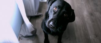
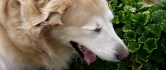
It is important to understand why dogs shake their heads. Shaking your dog's head and ears is a great way to get something out of the ears that shouldn't be there.
When dogs feel itching or irritation in their ears, they instinctively shake their heads. Although this may solve the problem if the dog has water in his ears, a piece of grass or an insect in his ear. But continued head shaking indicates that the irritation is ongoing and needs to be addressed.
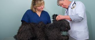
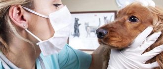
If your dog is constantly shaking his head and the behavior doesn't stop within a day or so, it's time to make an appointment with your veterinarian or try to fix the problem yourself.
Bacterial and yeast infections in the ear
The most commonly diagnosed health problem that causes dogs to shake their head and ears excessively is an ear infection.
Ear infections typically cause itching, excessive discharge, and inflammation, making your dog want to shake his head. If you lift your dog's ear and see redness, swelling, or discharge, there is likely an infection.
Ear mite infestations can cause similar symptoms, but they are not as common as yeast or bacterial infections in dogs (especially adult dogs).
Keep in mind that the infection can occur deep in your dog's ear, so the infection may be present even if you don't see obvious signs of it.
Otitis
Another possible disease is otitis media. This name hides a number of different inflammations of the hearing organs that appear for completely different reasons. This is a common and quite dangerous disease. If left untreated, the animal may lose its hearing forever.
The breeds most often susceptible to otitis media are:
- Labrador.
- Hunting.
- Setter.
- German Shepherd.
- French and English bulldogs.
- Shar Pei.
- Spaniels.
- Basset Hounds.
Some of the symptoms that appear coincide with previously described diseases, but there are also specific ones, by which it is easy to determine that this particular misfortune has overtaken the pet:
- Increased temperature of the affected area.
- Disease of only one ear.
- Unpleasant smell.
- Discharge. At first greasy and little different from sulfur, they gradually become a mixture of pus and blood and begin to leave abundant stains in all places where the dog lies or scratches.
You cannot cure otitis media on your own . The maximum you can achieve is to temporarily get rid of pronounced symptoms. But this will not help for long, the problem will return. The fact is that, in essence, otitis media is more a consequence than a root cause. For it to develop, a reason is needed. And until it is eliminated, the dog will not have healthy ears.
Most often the cause is:
- Injuries.
- Infections.
- Pathogenic bacteria.
- Fungal infection.
- Microscopic parasites.
If you suspect this disease, you must immediately contact a veterinary clinic to undergo a full diagnosis, identify the root cause and select an effective treatment.
Itching in the ear due to allergies
Allergies are another common problem that leads to head and ear shaking in dogs. For example, people may be allergic to ingredients in their food or to triggers in the environment (pollen, mold spores, dust or mites, etc.).
Allergy symptoms in dogs typically include a combination of itchy skin, hair loss, recurring skin and ear infections, persistent ear scratching, head shaking, paw chewing and muzzle rubbing on the ground.
Diagnosing a food allergy involves feeding the dog a diet that contains one carbohydrate (such as rice or potatoes) and one source of protein that has never been fed to the dog before (such as duck or venison) or that has been hydrolyzed (broken down into tiny, non-allergenic pieces).
The dog should eat only this food for a month or two. If symptoms disappear or at least improve significantly, a food allergy is likely.
Environmental allergies are best diagnosed using intradermal skin testing, but for some dogs a blood test is a reasonable option.
Diseases
There are several diseases in dogs that result in sudden head movements. Most of them are quite easy to cure, but there are also complex cases.
Otodectosis
Microscopic ear mite infestation is a common problem. It affects not only dogs that regularly go outside, but also pets that never go outside. An animal only needs to come into contact with a carrier once to become infected.
The main symptoms of such scabies are easy to recognize:
- The animal behaves restlessly and is constantly itching.
- Signs of infection are visible on both ears.
- Brown discharge with an unpleasant odor appears.
- When the skin is cleansed, microscopic wounds are visible on the affected area.
You can easily cure your dog of this type of parasite on your own. It is enough to keep the ears clean and regularly treat them with special preparations against parasites of this type and with antiseptics.
After all the symptoms disappear, it is necessary to carry out a full treatment with the drug against parasites and constantly update it.
Allergic reaction
Allergies are one of the most popular problems that cause dogs to feel discomfort in such a delicate organ. Determine that this particular problem has happened to your pet simply by additional signs:
- The tissues become red and slightly swollen.
- Small wounds and pimples appear.
- The combed surface becomes bald due to constant mechanical stress.
- The muzzle is swollen.
- My eyes are watering. Moreover, an opaque liquid may be released.
In order to stop an attack, it is enough to give a dose of an antihistamine. Choose a medicine specifically designed for dogs, and carefully calculate the dose based on your pet's weight. It is dangerous to use human drugs due to the possibility of overdose.
Antihistamines are only the first step on the path to recovery. It is also necessary to find out the cause of the incident and remove the allergen. Most often this is some new product or food component, but cosmetics or household chemicals, medicines or even a vaccine can also create problems.
If the symptoms do not disappear, then the allergen has not been found. To determine the problematic substance, you need to contact a veterinary clinic, where, with the help of a special study, they can determine what is causing such harm to the animal.
Water in ears
Head shaking, which occurs due to water getting into the ears, can be easily prevented by placing cotton balls (or half a cotton ball for small breeds) in the dog's ears before bathing or swimming.
Avoid pouring water on your dog's head while bathing. Instead, wash his body from neck to bottom and wipe his face and ears with a damp cloth.
If your dog doesn't tolerate cotton balls in his ears after swimming, consider wearing ear tape or cleaning his ears with a drying solution after swimming.
Your veterinarian can recommend a safe and effective product based on your dog's specific needs.
The dog shakes its head and scratches its ears
Our dogs may scratch their heads and ears throughout the day, and, especially in the summer, shake their heads to protect themselves from flying insects, and this is normal.
In the event that a dog begins to behave restlessly and at the same time scratches its ears and head too often and shakes its head, you should more carefully examine your pet’s head and ears for the presence of a particular disease.
The dog shakes its head usually from pain or itching.
Itching in a dog can appear on a wide variety of parts of the body, but the most common itching in dogs occurs in the ear area. During itching, the dog scratches its ears with its claws, rubs its head on various objects, scratches the skin at the site of itching until it bleeds, as a result, scabs form at this place.
The cause of itching in a dog can be:
Parasitic diseases. The most common cause of itching in dogs is the dog being infected by ectoparasites - fleas (fleas in dogs), lice (lice in dogs), mites (ticks in dogs).
The most common diseases in dogs are: Sarcoptosis canum (Sarcoptosis canum) is a parasitic itching disease of dogs caused by mites of the Sarcoptes canis species and clinically manifested in dogs by severe itching. The disease in dogs does not depend on the time of year. Dogs can be carriers of sarcoptic mange without showing clinical signs characteristic of the disease. Dog owners may have an allergic reaction to tick waste products - pseudoparasites.
Etiology. The causative agent of the disease (Sarcoptes Canis) is an intradermal parasite in dogs that parasitizes and multiplies in the epidermal layer of the skin.
Biological development cycle. The female lays 2-8 eggs in the epidermal layer of the skin, 40-60 in total. One generation of ticks develops under optimal conditions within 15-19 days, passing through the egg, larval, protonymph, teleonymph and imago phases. Male ticks live in the dog’s epidermis for up to 1 month, and die after copulation with the teleonympha. Female ticks live up to 1.5 months. Only teleonymphs and females infest healthy dogs. Once in the external environment, sarcoptic mange mites do not reproduce, but retain their mobility for up to 2 weeks and die at temperatures below 0°C. Eggs are viable for up to 1 month. Sarcotes in the dog's body feed on epidermal cells, lymph, and inflammatory exudate.
Pathogenesis. The dog's body is mainly affected in areas where there is no thick fur: ears, elbows and hocks. The disease in a dog begins to manifest itself 2 weeks after infection with a tick. Damage to the skin of a sick dog leads to disruption of the activity of many systems of the sick animal’s body (central nervous system, cardiovascular, reticuloendothelial, immune, etc. The dog’s skin becomes covered with papules, erythema, scabs, and even in some dogs there is superficial bleeding. In As a result of all this, the animal's skin respiration is impaired, oxygen deficiency increases, heat transfer increases.The dog's itching intensifies in a warm room, especially when it is near heating devices.
The course of sarcoptic mange can be complicated by allergic reactions to waste products secreted by mites. If timely treatment measures are not taken, the disease becomes chronic, which is characterized by lichenification, hyperpigmentation of the affected skin and hardening of the superficial lymph nodes. With reduced body resistance, irreversible pathological processes occur in a sick dog and the sick dog dies.
Clinical picture. At the early stage of the disease, the symptoms of sarcoptic mange are not pronounced. A week after a dog is infected with sarcoptic mange mites, during a clinical examination, a veterinarian diagnoses nodules on the bridge of the nose, brow ridges, and ears, which subsequently fill with clear liquid, turning into papules. The dog develops severe itching, as a result, scratches and scabs appear in place of the formed papules. The fur of such a sick animal is glued together with a liquid that is secreted by the affected skin. In the case of an atypical course of the disease, a veterinary specialist, during a clinical examination of such an animal, notes the appearance of a large amount of dandruff.
A veterinarian makes a diagnosis of sarcoptic mange based on anamnestic data (a survey of the dog owner), epizootic data, the clinical picture of the disease and confirms it with laboratory testing.
To detect the sarcoptic mite, its eggs or waste products, it is necessary to carry out deep scrapings of the affected skin. The most effective is taking scrapings near a fresh scratch, papules, in those places that a sick animal cannot lick. But even in this case, we can only detect a tick in 50% of cases.
Differential diagnosis. Sarcoptic mange must be differentiated from demodicosis (treatment and prevention of demodicosis in dogs), fungal skin diseases (trichophytia, microsporia, dermatophytosis), contact dermatitis, folliculitis and generalized pyoderma.
Treatment. Owners of a sick dog must isolate it from other dogs. Before treatment, the sick dog is thoroughly washed in warm water with tar or baby soap. For this purpose, the dog owner can use soap K, SK-9, and anti-soborrheic shampoo. During this procedure, the dog's skin is cleansed and freed from scabs, crusts, dandruff, fat, dirt, germs and other harmful deposits. After such a wash, the dog can be rinsed with infusions and decoctions of herbs - celandine, string, chamomile, calendula and others, then, in order to avoid colds and hypothermia, the dog must be thoroughly wiped with a clean rag. We shave the affected areas of the skin or cut them short. On wounds and scratches we apply anti-inflammatory and antimicrobial ointments, suspensions - Konkov, Vishnevsky, Wilkelson 1-2 times a day for 10-14 days, sulfur and sulfur-tar 2-3 times a day for 14 days or more, 1% liniment of chlorophos in fish oil, celestoderm, lorindene, flucenar, tetracycline, etc. Treatment of sarcoptic mange using the Demyanovich method using solutions of hyposulfite and hydrochloric acid is effective.
A 20% water-soap suspension of benzyl benzoate has a good antisarcoptosis effect. Skin treatment is repeated 2-3 times with an interval of 5-7 days. The method and use of these drugs in each specific case is determined by the attending veterinarian. After this, we begin to wash the sick dog in a bath with acaricidal preparations at intervals of 2 weeks. Good results in treatment are given by 0.5% emulsion of Shchudrin, decresyl, karbofos, 0.15% aqueous emulsion of diazinon, hexachlorane emulsion with 0.04-0.05% gamma isomer content. Treatment with acaricidal drugs for local skin lesions usually does not produce a positive effect. Due to the high toxicity of acaricidal preparations, puppies under 4 months of age cannot be subjected to this type of treatment. For young puppies, we treat each half of the body alternately. For focal sarcoptic mange in dogs, 2-3 times rubbing of a 0.5% oil solution of ASD-3 (Dorogov's Antiseptic Stimulator) is successfully used; the rubbing interval is 7-10 days.
Five-time treatments with Demos or Dekta with an interval of 3-4 days are effective. The sulfur contained in the drug helps restore the coat of a sick animal. Currently, for sarcoptic mange in dogs, drugs containing synthetic pyrethroids are most often used - stomazan, neostamazan, butox, ectomine, anometrine nitigifor, anometrine, permintin, ivomec, proposcur, etc. Based on these drugs, 0.1-0.2% are prepared aqueous solutions in which animals are bathed, and in case of focal lesions, the solutions are rubbed with a sponge or cotton swab into the animal’s fur and skin. After such treatment, the dogs are allowed to shake off and dry in a warm place, without washing or wiping. To prevent poisoning during treatment, the dog must be wearing a muzzle or special collars that prevent licking its fur. For the treatment of sarcoptic mange, the drugs Selamectin, Imectin are used - which are administered subcutaneously 2-3 times with an interval of one to two weeks and have side effects in some breeds of dogs - (Collie, Sheltie, Bobtail, Terriers, as well as dogs of dolichocephalic breeds). The drug Advocate, which combines imidacloprid 10% and maxidectin 2.5%, is very effective. This drug is produced by the Bayer company. At the beginning of treatment, if the itching intensifies in a sick dog, glucocortican is prescribed at a dose of 1 mg/kg daily for 3-4 days. When conducting a course of treatment with anti-scabies, veterinary specialists prescribe sedatives, antiallergic, antimicrobial drugs, as well as hepatoprotectors (Lif-52, etc.) in the treatment regimen for sarcoptic mange.
Treatment of the premises. If the treatment is carried out in winter, the dog’s things are treated with an anti-parasitic solution, boiled or sprayed with a 2% chlorophos solution, or even better, throw them away. It is imperative to disinfect the area where the dog’s bedding was located, as well as areas of the floor or rug where the animal usually lies. If treatment is carried out in the summer, then the dog’s things are thrown out into the sun. The tick disappears after 18 days.
Prevention. Owners separate and isolate dogs with sarcoptic mange from healthy ones and subject them to intensive treatment as prescribed by a veterinarian. Healthy dogs are treated with a variety of products that include acaricides. A variety of anti-parasitic collars are used.
Keep dogs clean (Dog Care) to prevent the appearance of endo and ectoparasites (ecto and endoparasites). Owners must organize adequate feeding of their animals (Approaches to feeding dogs, basics of feeding dogs, golden rules for rational feeding of dogs, feeding pregnant females, feeding lactating dogs, feeding puppies, feeding aging dogs). Have a monthly clinical examination at a nearby veterinary clinic. When contacting dogs with sarcoptic mange, you should carefully observe the rules of personal hygiene, since sarcoptic mange can spread to humans and cause scabies.
Demodicosis in dogs is a chronic parasitic disease of the skin of dogs caused by the microscopic worm-like mite Demodex canis.
Etiology . The causative agent of demodicosis in dogs is Demodex canis. Being itself an endoparasite, the mite is localized in the hair follicles and sebaceous glands.
Epizootological data . The following breeds of dogs are most susceptible to demodicosis: pugs, Charleys, Scotch terriers, French and English bulldogs. In terms of seasons, demodicosis is more widespread in the winter-spring period due to a decrease in skin tone in dogs, as a result of insufficient external insolation, which ultimately leads to the activation of mites and the clinical manifestation of the disease. Transmission of the causative agent of demodicosis (mite) occurs through direct contact with a dog with demodicosis, as well as through care items and clothing. Infection of puppies in dogs occurs in the first 3 months of life, in subsequent months of life due to keratinization of the epidermis of the skin, an increase in hair length, and difficulty in relocating ticks is observed. In the liquid exudate of pustules at a temperature of 17-20 degrees Celsius, the tick remains viable for 10 or more days, while on the walls and floor of an apartment at a temperature of 17-20 degrees it lives for only 20 minutes, and on a dog bed for about an hour. Freezing has a detrimental effect on the tick - it dies immediately. When heated to 50 degrees, it dies in 30-60 seconds.
The pathogenesis of demodicosis is associated with individual predisposition (when the normal physiology of the hair follicle is disrupted during molting, skin atony), as a result of which the mite easily penetrates the hair follicle. The course of pathogenesis is also influenced by the suppression of immunological reactions of the skin during hormonal disorders in dogs. Infection of healthy dogs occurs only through direct contact and only by sexually mature forms of the mite, which are selected from the affected hair follicles to the surface of the skin and actively move along it. More often, demodicosis lesions occur in those places where it is more elastic and where there are more folds and more humidity in the skin layer of air (head, chest).
The disease in dogs begins with the penetration of a tick into the hair follicle, which, using its paws, destroys the cells, the parasite penetrates deep into the follicle. Along the path of its movement, it feeds, gnawing out entire furrows (up to 70 microns) in the epithelium of the inner and outer root sheath. Moving to the excretory ducts of the sebaceous glands, the mite penetrates the sebaceous glands. Feeding, cutting off entire epithelial cells of the root sheaths, the mite begins to destroy the cells of the cortical layer of the hair root, as a result, the hair root becomes so thin that nothing remains of it and damaged hair falls out. Feeding in the sebaceous gland or at the bottom of the hair follicle, the mite makes numerous damage to their epithelial layer. At the same time, other mites also penetrate into the same follicle. Reproducing in the follicle, they form entire colonies of ticks (up to 200), in which there are heterosexual individuals of all stages of the biological cycle (larvae, nymphs, adults). In a sick dog, a demodectic focus thus forms in the skin, into which pyogenic microflora then penetrates. (staphylococci, etc.).
In generalized demodicosis, hereditary T-cell deficiency plays a role. With long-term demodicosis (pyodermodecosis), the function of the dog's liver and gastrointestinal tract is impaired.
Clinical picture. Demodicosis in dogs is characterized by general clinical symptoms: loss of appetite, depressed state of the sick animal, itching, redness of certain areas of the skin, formation of scabs, nodules on the skin and the inner surface of the ears.
- Scaly (squamous) form of demodicosis. During a clinical examination of dogs with demodicosis, a veterinarian finds round, hairless areas of skin on the brow ridges, nose, forehead, lips and limbs. As a result of constant hair loss, the veterinarian visually sees round, bald areas of skin in a sick dog, which can sometimes be clearly demarcated. When examining these lesions, the veterinarian notes slight redness of the skin, a large number of pityriasis-like scales, the skin in this area is rough, cracked on palpation, sometimes with small nodules. The hair at the edges of the demodicosis lesion is weakly strengthened, unevenly distributed and brittle. At a later stage of the disease, the skin becomes grayish-bluish with rounded redness. In the squamous form of demodicosis in dogs, the ventral and dorsal surface of the skin is affected.
- The pustular form (pyodemodecosis) can develop either from the squamous form or independently. Upon clinical examination, the veterinarian will notice swollen and reddened skin of a sick dog, with small, hard nodules located next to the hair follicles and having a blue-red hue. Subsequently, these nodules quickly turn into yellow, red-brown, and in some sick dogs, black pustules. When pressed with fingers, greasy pus is released from the abscess, sometimes mixed with blood, in which mites can be found at all stages of the life cycle. When a secondary infection penetrates into the demodectic focus of a sick dog, we register extensive pyoderma with the formation of ulcerative abscesses. The affected areas of the skin are thick, wrinkled and moist, and some of the skin is cracked. During a clinical examination, the dog exhibits severe itching. An unpleasant odor emanates from the affected skin area. With pyodemodecosis, we palpate enlarged and painful submandibular lymph nodes; in some sick dogs we note lameness and purulent phlebitis. English and American Cocker Spaniels sometimes have demodicosis of the paws. Clinically manifested by hair loss, cellulite, erythema, furunculosis, and in severe cases, severe lameness and purulent phlebitis of the veins. They also have otodemodectosis, which is clinically manifested by hyperemia of the inner ears, which are painful and hot on palpation. When scraping from the affected areas of the skin, we find many mites at different stages of development. In such dogs, there is abundant formation of wax in the ears and the appearance of crusts. In some dogs, a generalized form of demodicosis occurs.
A veterinarian makes a diagnosis Scrapings must be taken from several affected areas, penetrating deeply into the skin (until blood appears). In the pustular form, we examine the pustular liquid by applying it in drops to the glass.
Differential diagnosis . Demodecosis must be differentiated from autoimmune dermatosis, eczema, sarcoptic mange, staphylococcosis of dogs and cats, alopecia of endocrine nature, fungal skin diseases (trichophytosis, microsporia, dermatophytosis), pyoderma of bacterial origin, bacterial furunculosis.
Treatment . Demodicosis belongs to the group of skin diseases of dogs that are difficult to treat. It can be especially difficult to treat generalized pyodemodecosis, due to the involvement of the dog’s entire body in the pathological process. The difficulties of treatment lie in the difficulty of delivering the active substance to the location of the ticks (in the tick colony) in order to completely destroy them.
When carrying out complex treatment of demodicosis, the veterinarian must take into account that systemic acaricides (organophosphorus preparations, ivermectins, pyrethroids, etc.) kill the adult tick, while the preimaginal stages (which are in a passive state) do not die because they don't eat anything. After stopping treatment of a sick dog, the larvae of the nymphs again enter an active state, begin to reproduce, and the number of demodectic mites is quickly restored. Therefore, when starting complex treatment of demodicosis, a veterinarian must conduct treatment based on suppressing the vital activity of D. Canis mites. Having started complex treatment, it is necessary to exclude all predisposing factors, not to use corticosteroid drugs, and treat secondary pyoderma with systemically acting drugs (after determining sensitivity in a veterinary laboratory). During treatment, every 3-4 weeks it is necessary to take control skin scrapings and examine them for the presence of mites. Treatment of a sick dog is stopped only when 3 negative results of control skin scrapings for demodicosis are obtained.
When treating demodicosis in dogs, the following medications are used: - 1% trypansini solution in physiological sodium chloride solution. The technique for its use is as follows: add the appropriate amount of trypansini to a hot (80-90 degree) physiological solution of sodium chloride, filter and sterilize in a water bath for 30 minutes from the moment of boiling. The cooled solution is administered intravenously to a sick dog at a dose of 0.5-1.0 ml per 1 kg of body weight. A 1% trypansini solution must be administered to a sick dog at least 4 times with an interval of 7 days.
Recently, veterinary clinics have been using the imported drug berynyl (Germany). It is administered to a sick animal subcutaneously in the form of a 7% solution at a dose of 3.5 ml/kg of animal body weight 3 times with an interval of 16 days. Before using these medications, sick dogs must take cardiac medications (camphor oil, sulfacamphocaine, caffeine, etc.). Of the organophosphate preparations in veterinary clinics for the treatment of demodicosis in dogs, chlorophos (trichlorphan, neguvan), sebacil ronnel, and safely are used. Every other day we wash the entire surface of the body with a 2% solution of chlorophos until complete recovery (disadvantage - it does not have a systemic effect and does not penetrate the demodicosis focus). Ronnel - dissolved in propylene glycol (180 ml of 33% Ronnel per 1 liter of propylene glycol) applied daily to the affected area until recovery (6-10 times). If you have to treat more than 1/3 of the body surface, atropine sulfate, phospholithin or dipyroxime are used to relieve the resulting toxicosis. Glikhlovos (dematef) - treat the affected area; in the generalized form, the drug must be applied along the spinal column, retreating 2-3 cm at a dose of 0.17 ml per 1 kg of body weight, 4 times with an interval of 7 days. Sayfli (cythioate) – 1 tablet per 10 kg of body weight 2 times a week for 6 weeks. For the squamous form, soap K is used. It is used in the form of a 5% aqueous emulsion, generously moistening the affected area, 6-8 times with an interval of 5 days. Pyrethroid drugs: Pedims, Cybok, Nanacid, Cydem. Pedems - applied to the affected area at the rate of 1-1.5 ml per 1 kg of body weight, 2 times, an interval of 7 days. Cydem - in aerosol and propellant-free cans - is applied to the skin at a distance of 5-10 cm in a dose of 1 g per 1 kg of body weight. Treat 4 times at an interval of 7 days. Deces, Danitol, Baytikhol - in the form of oil solutions in 0.025% concentration 3-4 times with an interval of 10 days by rubbing into the affected area of the skin.
Recently, canine demodicosis has been treated with ivermectin drugs. Ivomec – subcutaneously at a dose of 250 mg per 1 kg of body weight with an interval of 6-7 days. We do 2-6 injections. Ivermectin is administered orally at a dose of 0.6 mg/kg daily for 2 weeks. When treating generalized demodicosis, Vaganova ointment is used (ASD -3 fraction -100.0, sulfur - 100.0, birch tar -20.0; Lysol -30.0, petroleum jelly -800.0). The course of treatment is up to 1 month. For generalized demodicosis, amitrosis is used. The French company produces collars for dogs that contain amitrose. This collar is changed once a month. The course of treatment is 3-4 months. The effectiveness of treatment with amitrosis for generalized demodicosis increases when it is used with immunomodulators (immunofan, ribotan, fakrinil, gamavit). A good effect can be obtained from autohemotherapy. The ears of dogs are treated with aerosols - acrodex, dermatosol, cyodrine, psorotol, perod from a distance of 10 cm by pressing the valve of the aerosol can for 1-2 seconds. For any form of demodicosis, the use of Gamabiol balm is indicated, which relieves inflammation of the skin and subcutaneous tissue.
Pathogenetic therapy. For better hair and fur growth, as well as to relieve swelling of the skin, owners of a sick animal are advised to add pure sulfur to their food, as well as rub liniments containing sulfur into bald areas. When skin resistance is suppressed, sick animals are prescribed thyroidine, and 1-2 drops of iodine tincture (5%) must be added to the diet. Symptomatic therapy. At the recovery stage, Fir and Sea Buckthorn oil, as well as an oil solution of vitamin A, are used to soften the skin and stimulate regenerative processes in the skin. In case of complications with staphylococcosis, treatment with antibiotics (subtitrated). When treating demodicosis, vitamin preparations are used: pushnovit, gendevit, vitamins according to Ryss, etc. The effectiveness of the treatment is checked after 25, 30 and 45 days. It is mandatory to do skin scrapings and remove an acarogram.
Prevention. Dog owners must provide adequate feeding for their pets. Have a monthly clinical examination at a designated veterinary clinic. Regularly clean and wash with hot water (60-70 degrees) the dog’s cage, rugs, and resting place. In order to prevent demodicosis in dogs, it is necessary to exclude the development of immunosuppression in puppies. Do not use corticoids for up to 1 year. Properly and timely deworming of dogs. Pet owners should ensure that the dog's fur is not wet. Keep the dog clean (Dog Care), prevent the appearance of endo and ectoparasites (ecto and endoparasites). A good preventive measure is the use of insecticide collars. Prevention of puppies from infection with demodicosis is carried out by ectoparasitic and endoparasites by treating puppy bitches with the drug Ivomec at a dose of 200 µg/kg. This treatment is done 6-7 days before whelping.
The cause of itching in a dog can be fungal diseases - microsporia, trichophytosis, dermatophytosis.
Bacterial infections – Streptococcosis (streptococcosis of dogs and cats), staphylococcal infection in dogs.
Food and non-food allergies . Today, food allergies, especially in purebred dogs, are quite common. For more information about food allergies, see our article on food allergies in animals.
Stressful situation . Itching in a dog can appear as a result of severe stress, especially if the dog lacks proper attention from its owner.
Your dog may experience short-term itching as a result of numerous mosquito bites while walking. It is necessary to keep in mind that simultaneously with a mosquito bite, your dog can get such a dangerous disease as dirofilariasis.
Clinical picture. The main symptom that provokes the appearance of itching in a dog is the presence of various skin lesions in different parts of the body, the presence of a pink or reddish tint at the site of the itching, the affected area of skin is usually painful and moist upon palpation, and we note an increase in local body temperature. Change in the dog’s behavior – the dog becomes restless, actively scratches the ear area with its paws, rubs its muzzle and eyes on various objects, scratches the perineal area, etc. in the head and neck area.
Diagnosis of the causes of itching. The only way to accurately determine the cause of your dog's itching is to visit a veterinary clinic.
One of the reasons why a dog shakes its head and scratches its ears may be otitis (ear disease in dogs).
A dog may shake its head as a result of an injury to the head.
When foreign bodies or water get into the ears, the dog begins to shake its head to get rid of them.
Serious conditions associated with head shaking
Other health conditions that can cause dogs to shake their heads excessively include foreign objects lodged in the ear canal, inflammatory diseases, or even neurological disorders that cause head tremors, which are easily confused with head shaking.
If your dog has recurring ear infections, you and your veterinarian will need to find the underlying cause, such as allergies, anatomical abnormalities, or hypothyroidism.
Diagnosing and treating the cause of your dog's head and ear shaking is important not only because it is a symptom of a potentially serious problem, but also because prolonged or particularly severe head shaking can rupture blood vessels in your dog's ear.
The ear hematomas that result often require surgery to treat, so whenever possible we should prevent excess head shaking and not only treat it when it develops.
Treatment with folk remedies
Non-traditional treatment methods are used not only in the initial stages of the development of various ear diseases in dogs, as a complement to the main therapy, but also for preventive purposes.
Treatment with folk remedies alleviates the condition of animals and speeds up recovery.
- The use of decoctions of medicinal plants that have antibacterial, anti-inflammatory, wound-healing effects.
- For washing and treating the ears, soda solutions, alcohol tinctures (propolis tincture), vegetable oils (camphor), strong black tea, solutions, liniments prepared according to special recipes are used.
Remember! Non-traditional methods of treating ear diseases are effective only in the early stages of their development. Before treating your pet with folk remedies, consult your veterinarian.
Causes
Anatomical features predispose to the development of pathological processes in the hearing organs. Long-haired dogs with large floppy ears are most often affected. Ear wax accumulates in them and creates a favorable environment for the development of pathogens of infectious and non-infectious diseases. The following are the causes of ear problems in dogs:
- foreign particles;
- injuries;
- ear mite;
- fungal infection;
- neoplasms;
- allergic conditions.
Long-eared animals are prone to otitis media
Foreign particles
During hunting or walking, seeds and vegetative parts of plants get into the ear, which bother the dog. When swimming, the ear canal becomes clogged with water.
Injuries
During fights between aggressive dogs, hematomas occur. If there is no inflammation, they are not dangerous, but cause discomfort, manifested by shaking of the ears.
Ear mite
Parasites constantly live on the inner surface of the shell. The reproduction of microscopic arthropods is controlled by the immune system. When it weakens, the parasite strengthens and actively feeds on tissues. The meal is joined by another permanent resident - the yeast fungus Malassezia. The waste products of pathogens cause an allergic response.
Otodectosis
Fungal infection
It is the result of activation of micromycetes during otodectosis, blockage of the auditory canal with earwax, particles of wool, and foreign objects.
Neoplasms
The pathology can be congenital or developed as a result of an inflammatory process caused by hormonal disorders.
Malignant neoplasm
Allergic conditions
Dogs show an inadequate reaction to perfumes, dietary components, dust, chocolate, and other delicacies intended for people. Allergies can be a consequence of exposure to cutaneous and intradermal parasites, as well as side effects of medications.
Allergic otitis media
Information for dog owners.
Otodectosis in dogs is extremely rare. Treatment of otodectosis in dogs is no different from that described for cats, using drugs as “Bars Forte ear drops for dogs” and “Aurizon” ear drops for dogs. We discussed the first drops in detail earlier. instructions, as well as the price of Aurizon ear drops, on specialized websites. With timely detection and treatment of otodectosis, the prognosis is favorable.
And in order for your pet to always be healthy and not suffer from ear diseases, do not forget about the basic preventive measures:
- ear hygiene,
- regular treatment of your pet with antiparasitic drugs,
- balanced nutrition and preventive examination.
Bruises on ears
These bruises are not only caused by inflammation of the blood vessels, but any of the mentioned reasons due to which the dog often shakes its head from side to side will lead to bruising if the ears are hit during this process.
© shutterstock
These bruises are also only visible if the blood vessels have ruptured due to the shaking, further exacerbating the problem as this inflammation adds to the overall discomfort the dog is already experiencing. To stop the deterioration, the underlying cause of the discomfort must be addressed.
Diagnostics
Only a veterinarian can make a diagnosis based on the information collected and laboratory tests. The owner should be alert to symptoms that bother the pet:
- whines in pain, rubs his ear with his paws;
- when shaking your head, squelching sounds are heard;
- swelling, redness of the skin;
- unpleasant odor from the ears;
- enlargement of the submandibular lymph nodes;
- lethargy, apathy, depression.
To understand what to do when a dog shakes its ears, you need to find out the reasons that caused this behavior.
How to properly care for dog ears
Dog ears come in different shapes, and this requires different approaches to caring for them:
- hanging like a basset hound;
- erect, like a German Shepherd - ideal, requiring no maintenance;
- pink, folding like a bulldog's;
- semicircular, erect, shaggy, like a collie's.
Those with floppy ears suffer from excess moisture - especially if they are waterfowl dogs. They have a harder time drying the skin around the ear and increase the risk of bacterial contamination.
Did you know? Dogs hear 4 times better than humans.
Breeds with folds and folded ears need to wipe the interfold space and prevent the development of yeast fungi in it.
Ear care rules:
- Check them weekly for signs of inflammation or signs of mites.
- During the test, smell the surface of the ear. A healthy ear should not smell.
- After inspection, clean with special solutions.
- Wipe the folds with an antiseptic for breeds such as Shar Pei or Bulldog.
How to tell if your dog's ears are itchy
If a dog scratches its ears with its paw, then they probably itch, or rather, they itch very much. This is accompanied by skin irritation - it becomes inflamed, red and swollen. And if the pet scratches them very actively, then scratches will be added to the inflammation, through which the infection gets inside, causing complications. Only occasional, infrequent scratching is normal. In this case, there should be no symptoms of diseases, external parasites, fungi or bacteria.
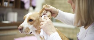
First aid:
- Take a flashlight and look inside your ear canal. A smooth, shiny surface with a delicate pink tint means there are no problems.
- If this is not the case, proceed to cleaning.
- Some dogs may require hair removal that grows in the outer ear canal.
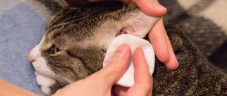
For cleaning you need cotton swabs and cleaner. Popular products: Trixie lotion, 8in1 Pro-Sense Ear Cleanser lotion, Auricap spray, Espree Ear Care. Almost all modern drugs have antibacterial and antifungal properties, and also perfectly remove wax and protect the ears from inflammation.
Cotton swabs should be used to clean the ribs of the outer ear, but should not be inserted into the ear canal to avoid damaging it.
Proper cleaning consists of the following steps:
- Hold your ear upright and gently drip the cleanser into the ear canal.
- Massage for 20 seconds, then release and allow the dog to shake its head, distributing the product into the ear canal if necessary. Massage helps soften the contents.
- Using a cotton swab, gently wipe the visible part of the ear canal and outer ear.
- Reward your pet with a treat and move on to the second ear.
If you see that the ear canal is inflamed or there are other signs of a problem, then postpone cleaning. The ear in the “as is” state is much easier to examine and diagnose, since nothing interferes with this. A pure hearing organ will not provide the doctor with such a volume of valuable information.
Important! Avoid using alcohol-based ear cleaners. They irritate sensitive or inflamed skin.
Prevention
The best way to combat a disease is its prevention. To prevent your dog from getting otitis due to parasitic diseases, treat your pet regularly for fleas and ticks. Drugs such as Simparica and Stronghold are active against these parasites and will protect your pets from uninvited guests.
Keep an eye on your pet's ears by cleaning them regularly with lotion and a cotton pad. Do not clean your ear with cotton swabs - this can cause harm. To clean your dog's ears, pour a small amount of lotion into the ear canal, massage the base of the ear and allow the dog to shake its head vigorously. Excess wax will end up on the inside of the ear, where it can be easily wiped off with a cotton pad or gauze.
If you know that your dog is prone to food allergies, carefully follow the diet and talk with all family members so that they do not feed it from the table. This is extremely important for allergic animals. For other types of allergies, when the first signs of exacerbation appear, contact your veterinarian as soon as possible to prevent scratching.
Health to you and your pets!