11/14/2021 19,146 Interesting facts about dogs
Author: Olga
Surely every dog breeder or simply a fan of man’s four-legged friends will be interested in learning about what the “internal structure” of dogs is like? What do we and our pets have in common, and how are we strikingly different? Therefore, we suggest making a detailed excursion into the world of canine anatomy right now!
[Hide]
Skeletal structure
Naturally, studying the anatomy of any animal begins with studying the structure of its skeleton. The skeleton of a dog is the foundation, the frame that holds all the organs and muscles of the dog inside. Let's look at all the “components” of a dog’s skeleton one by one.
Scull
The skull of dogs is usually divided into facial and brain parts. Both of these parts consist of paired and unpaired bones (discussed in the table below).
| Paired skull bones | Unpaired bones |
| Frontal | Interparietal |
| Parietal | Lattice |
| Temporal | Wedge-shaped |
| Reztsovaya | Sublingual |
| Lower jaw | Opener |
| Nasal | Pterygoid |
| Palatal | Occipital |
| Zygomatic | |
| Maxillary | |
| Tearful |
It is easy to calculate that a dog’s skull will consist of 27 bones, which are securely connected to each other by connective cartilage tissue. As the dog gets older, this tissue ossifies. In this case, the lower jaw is attached to the skull with the help of a strong movable joint, which allows the dog to chew food.
Note that the shape of the skull of dogs can vary greatly. In the process of selection, people contributed to the fact that some breeds were recognizable precisely due to the original structure of the skull.
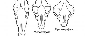
Thus, according to the shape of the skull, dogs are divided into long-faced, short-headed and dogs with normal head length. Moreover, it is the facial part of the skull that will make the biggest differences. The general name for all breeds with a shortened facial part of the skull is brachycephalic.
Vivid examples of the brachycephalic structure of the skull are Pekingese, Bulldogs, Pugs, Boxers, and Shar-Peis. These dogs have a wide parietal part of the skull, a greatly shortened and flattened facial part and a protruding jaw. This special structure is the result of many years of selective breeding work, when individuals with the desired trait, in this case a flattened muzzle, were deliberately selected. However, such an unusual symptom turned out to be associated with significant health problems.
After all, the disproportionately short muzzle caused degenerative changes in the structure of the dog’s respiratory tract. Because of this, all of the above breeds are prone to tracheal collapse, pulmonary hypertension and excessive tear production. Surely everyone has noticed that outwardly cute Pekingese or Pugs often walk around “tear-stained”, and every breath they take is accompanied by wheezing or grunting. To describe all the inconveniences that a brachycephalic dog experiences, there is even a special term - brachycephalic syndrome.
However, let's return to the structure of the skull and say a few more words about the teeth and bite of the dog. Thus, the dental system of dogs requires the presence of canines, incisors, molars and premolars. An adult dog should have 42 teeth, and the baby jaw consists of 28 teeth. The bite of dogs can be different, it depends on the breed and the standard prescribed by this breed.
There are the following types of dog bites:
- Scissor-shaped, when the upper incisors in a closed form cover the lower ones. In this case, the lower incisors are closely adjacent to the upper ones.
- Pincer-shaped, the incisors of both jaws adjoin each other with a cutting surface.
- Undershot, the lower jaw is inferior in length to the upper, so there is free space between the dog’s incisors.
- An undershot jaw, the lower jaw protrudes forward, is also called a “bulldog” jaw.
Torso
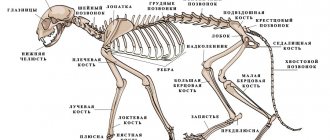
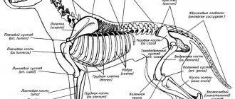
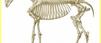
The dog’s body itself will consist of the spinal column - the axis of the body and the ribs that are attached to it and together make up the dog’s skeleton (in the picture below you can see the dog’s skeleton).
The dog's spine, in turn, consists of the following sections:
- cervical - formed by seven vertebrae, the first two are more mobile and are called atlas and epistropheus, like in cats;
- thoracic - made up of 13 vertebrae;
- The lumbar region, like the cervical region, consists of 7 vertebrae;
- The spinal column is completed by the sacral section, the single sacral bone of which is made up of 3 fused vertebrae.
The tail consists of 20-23 movable vertebrae. The chest is represented by 13 pairs of ribs, 9 of which are true and attached to the sternum, and 4 false form the costal arch. Dogs' ribs provide reliable protection for the heart and lungs and have different curves depending on the breed. The lumbar vertebrae are large and have many spurs, thanks to which the muscles and tendons that hold the abdominal organs are securely attached to them. The sacral vertebrae fuse into a single strong bone, which serves as a transition between the loin and the tail.
The first five vertebrae of the caudal region are the most developed and mobile. According to the standard of some breeds, the caudal vertebrae are docked in the quantity prescribed by this standard.
Limbs
The limbs of dogs have a rather complex structure. The forelimbs are a continuation of the obliquely set scapula, which passes into the humerus with the help of the glenohumeral joint. Next comes the forearm, where the radius and ulna bones are connected by the elbow joint. This is followed by the carpal joint, which consists of 7 bones connected to the 5 bones of the metacarpus.
The metacarpus consists of 5 fingers, 4 of them have three phalanges, and 1 has two. All fingers are “equipped” with claws, which, compared to cats, are not retractable and consist of strong keratinized tissue.
The front legs are attached to the spine by strong shoulder muscles. Due to the fact that the upper parts of the shoulder blades protrude beyond the thoracic vertebrae in dogs, a withers is formed - an indicator of the dog’s height. The hind limbs are represented by the femur and tibia, where the connecting elements are the hip and knee joints.
The lower leg, which consists of the tibia and fibula, is attached to the tarsus using the hock joint. The tarsus, in turn, passes into the metatarsus and ends in 4 fingers with three phalanges. A detailed explanation of the structure of the dog's foot is available in the video below.
Dog spine: anatomy
The spine consists of the following bones:
- 7 – cervical
- 13 – thoracic vertebrae
- 7 – lumbar
- 3 – sacral
- 20-23 – tail
The first cervical vertebra is called the atlas, and the second is called the epistropheus. Both of them are somewhat different in structure from the rest of the vertebrae of the dog's column. Thanks to them, the mobility of the animal's head is ensured. The ribs are attached to the thoracic vertebrae through the joints, which form the dog's chest.
The tail vertebrae are very mobile, but in some dogs, according to the standard, the tail must be docked.
Internal organs
Naturally, familiarity with the anatomy of a dog cannot be limited only to the skeleton and musculoskeletal system. If we already have some idea about the skeleton of a dog, let's talk about its internal organs and systems.
Digestive system
The digestive system of dogs is very similar to the digestive system of other mammals, including you and me. It begins with the oral cavity, which is equipped with strong and sharp teeth. Our pets are predatory animals, and therefore their jaws are adapted to eating large pieces of meat. Moreover, food is not always crushed in the mouth; dogs often swallow fairly large pieces whole. Our pets begin to actively produce saliva from the mere smell of food and its appearance, and the enzyme composition of saliva is slightly different; each breed has its own.
The food then moves through the esophagus and reaches the stomach. The main “digestion” occurs in this muscular organ. Gastric juice and special enzymes, under the influence of peristaltic processes, transform food into a homogeneous mass called chyme. At the same time, the stomach valves should not allow food to return back into the esophagus or enter the small intestine prematurely. At least this is how a healthy dog's digestion should proceed.
Well, the small intestine, which is next in line, closely “interacts” with the pancreas, duodenum and liver. Pancreatic and gallbladder enzymes continue to act on the chyme. And the walls of the small intestine actively absorb useful substances from it in order to “transmit” them into the blood. At the same time, the small intestine is quite long, and its absorption area is impressive - depending on the breed, it can be equal to the area of a room!
The digested food then moves into the large intestine. By this point, all the beneficial substances have already been taken from it, only water and coarse fiber can remain. Feces will be formed from the remains of waste food, water, some bacteria and inorganic substances. Defecation occurs under the control of the central nervous system; in case of nervous disorders or old age, bowel movements may be uncontrollable.
Respiratory system
The dog's respiratory system performs a vital function: thanks to it, all cells of the body receive the required dose of oxygen, and waste carbon dioxide is removed. The respiratory system of all mammals, and dogs are no exception, is usually divided into upper and lower sections. The upper section consists of the nasal cavity, nasopharynx, trachea and larynx. The movement of air begins through the nasal passages - nostrils, the shape and size of which depend on the breed of the dog. In the nasopharynx, the inhaled air is warmed, and thanks to the nasal glands, the air is “filtered” from dirt and dust.
Next, the air moves through the larynx, a cartilaginous organ that is held by the hyoid bone and is equipped with vocal cords, that is, it is responsible for sound production. Next comes the trachea, also a cartilaginous organ, closed by the tracheal muscle. The lower part of the respiratory system is represented by the lungs and bronchi. The lungs, in turn, consist of 7 lobes and are heavily dotted with blood vessels to enrich them with oxygen. The lungs are an organ that can significantly change its volume: when you inhale, they increase many times over, and when you exhale, they seem to “deflate”.
This elasticity is possible thanks to the rhythmic contractions of the diaphragm and intercostal muscles. During inhalation, old air is “replaced” with oxygen-saturated new air in the alveoli of the lungs. The respiratory rate of dogs should be in the range of 10-30 breaths per minute, it depends on the breed and physical condition of the pet. Small dogs breathe more frequently than large dogs. The breathing rate can change greatly in case of fear, heat and physical exertion.
Circulatory system
Naturally, the main organ of the circulatory system is the heart. Through arteries, blood is distributed to all other organs, and through veins it returns to the heart. The dog's heart is a strong muscular hollow organ that is located between the 3rd and 6th ribs in front of the diaphragm.
The heart has four chambers and is divided into two parts: right and left. Both parts of the heart are in turn divided into the atrium and ventricle. In the left side, arterial blood circulates, entering there through the pulmonary veins, in the right - venous blood, which enters the heart from the vena cava. From the left side, oxygenated arterial blood flows into the aorta.
The heart provides a continuous flow of blood in the body, it moves from the atria to the ventricles and from there enters the arterial vessels.
In this case, the walls of the heart consist of the following membranes: the inner membrane - the endocardium, the outer membrane - the epicardium and the cardiac muscle of the myocardium. In addition, the heart has a valve apparatus, which is designed to “monitor” the direction of blood flow and to ensure that arterial and venous blood does not mix. The size of the heart and the frequency of its contractions greatly depend on the breed of the dog, its gender and age, and environmental factors.
The first indicator of a dog's heart function is the measurement of the pulse, which normally ranges from 70-120 beats per minute. Young individuals are characterized by more frequent contractions of the heart muscle. The complex device has a system of capillaries and blood vessels of the dog, which literally “penetrates” the entire body of the animal and all its organs. For 1 sq. mm of tissue there are more than 2500 capillaries. And the total volume of blood in a dog’s body is 6-13% of body weight.
Excretory system
The excretory system of our little brothers cannot function without internal organs such as kidneys (available in duplicate). They communicate with the bladder through the ureters and end in the urethra. The purpose of the excretory system is the formation, accumulation and removal of urine from the animal’s body. Through urine, the body is freed from metabolic products; any violations in this process are fraught with serious health problems, including death.
To filter blood, the kidneys are equipped with nephrons, each of the nephrons is enveloped in a network of tiny blood vessels. As an animal ages, nephrons will break down and be replaced by scar tissue, making kidney problems common in older animals.
Reproductive system
The reproductive system is closely connected with the excretory system. Anatomically, in males, the urinary canal is also the vas deferens; in addition, for reproduction, males need testes and an external genital organ. In this case, in a newborn male dog, the testicles are located in the abdominal cavity, but by two months they will descend and take their place in the scrotum. It is there that the sperm will subsequently “mature”. In addition to the testes, males have a prostate, a sex gland that maintains the viability of sperm.
The male penis, consisting of a head, body and root, is covered by a preputial sac; at the moment of arousal, the sexual organ comes out of the sac and this is called an erection. Moreover, the hardness of the penis is achieved not only due to the cavernous bodies, but also due to the bone located at the base of the organ. Puberty in males, as well as in females, occurs at 6-11 months; small dogs “mature” faster. But males are allowed to mate at 15-16 months, and females at 1.5-2 years; by this age, dogs have completely completed puberty and will definitely give birth to healthy offspring.
The genital organs of females are the uterus; by the way, the uterus of dogs has “horns” to which the ovaries, fallopian tubes and vagina are “attached”. The egg of a female dog, like that of a human, matures in the ovaries. This process is quite complex and occurs under the constant “control” of hormones. As estrus approaches, the follicles containing the egg enlarge, and when estrus occurs, the follicle bursts, clearing the way for the egg. The egg matures in the fallopian tubes for another three days, while the fluid from the ruptured follicle produces a hormone that prepares the female’s body for pregnancy.
Bitches come into estrus twice a year, but dogs of northern breeds have estrus once a year and it lasts about 28 days. The optimal time for mating is 9-14 days of estrus. If a female mated with two males, then her litter may contain puppies from both males. Therefore, breeding of purebred dogs always occurs under close control from the owner. And one more nuance: dog embryos do not develop in the uterine cavity, but in the horns - tube-shaped processes on both sides of the main reproductive organ.
Nervous system
The nervous system of dogs is represented by central and peripheral sections. The central nervous system consists of the brain and the spinal cord adjacent to it, and the peripheral nervous system consists of many nerve endings and fibers that penetrate all the organs and tissues of the animal. Bundles of nerve fibers make up nerve trunks, which are more simply called nerves. All nerves are divided into afferent and efferent. The former transmit “information” from the organs to the control center - the brain, and the latter, on the contrary, transmit impulses arising in the brain to the organs and tissues of the dog.
The building block of the dog’s entire nervous system is the nerve cell, which necessarily has processes. The transmission of nerve impulses occurs through the contact of nerve cell processes and with the help of mediators. Mediators are substances that transmit impulses. Information is transmitted through nerve cells and fibers as if by telegraph, and the transmission speed is about 60 m/s.
Briefly about the main thing
- The skeleton consists of 290 bones, 262 joints. It supports the body and protects the organs.
- The jaw is one of the parts of the skull. An adult dog should have 42 teeth, and the bite depends on the breed.
- The internal organs are formed into the digestive, respiratory, circulatory, urinary, nervous and reproductive systems.
- Dogs' sense organs (with the exception of taste) are much better developed than those of humans. The main elements are eyes, ears, nose, tongue.
Do you know the location of your dog’s internal organs well? Share interesting facts about the structural features of these animals in the comments.
Sense organs
Dogs' sense organs are extremely developed. This predator is able to hear and smell much better than you and I. Therefore, we propose to talk about the dog’s senses in more detail, because without them the dog would not be the same as we are used to seeing it.
Structure of the eye
The eye of our four-legged friend consists of three membranes: fibrous, vascular and retinal. In principle, the structure of a dog's eye is anatomically very similar to our organ of vision. The principle of perception of visual information in a dog is no different from the principle of perception of all other mammals. A beam of light passes through the cornea and hits the lens, which focuses the light onto the retina, on which the light-perceiving elements are located. The light-perceiving elements in dogs, just like in us, are rods and cones.
The human eye is equipped with a so-called yellow spot - the place of the highest concentration of light-receiving elements; dogs do not have a yellow spot, so their vision is worse than that of humans. However, a dog can perceive information better in different lighting conditions, so our friends navigate in the dark much better than us.
Ear structure
Our four-legged pets perceive a lot of information through hearing, which they have much more acutely than you and I. The dog's auditory analyzer begins with the outer ear, moves to the middle ear and ends with the inner ear. The outer ear begins with the auricle, which is necessary for capturing sounds and directing them to the deep parts of the auditory organ. The auricle is a cartilaginous organ to which muscles are attached, allowing it to be rotated to improve focusing on the source of sound. The external auditory canal follows the auricle and is divided into horizontal and vertical parts.
Essentially, the ear canal is a skin tube through which sound travels to the eardrum. The skin of the auditory canal contains numerous glands, and hair often grows profusely in the auditory canal of dogs. Next comes the eardrum - the thinnest membrane, it serves to separate the outer and middle ear and capture vibrations of sound waves. The middle ear can be characterized as a bony cavity, which is the “receptacle” of the auditory ossicles (hammer, stapes and incus) and the inner ear. The auditory ossicles are attached to the inside of the eardrum and greatly amplify sound vibrations, transmitting them to the structures of the inner ear.
The inner ear is a container for auditory receptors and an organ of balance - the vestibular apparatus. It is in the inner ear that sound vibrations are analyzed and information is generated for transmission to the brain.
Structure of the nose
A dog’s nose is a hypersensitive organ; in principle, we can say that our four-legged friends live in a world of smells. Animals associate everything that surrounds them with some kind of smell, including you and me. A dog's nose is equipped with 125 million olfactory receptors, while our humble nose has only 5 million. The mucus that covers the inner surface of both our and the dog's nose, in dogs extends beyond the olfactory organ and also covers its outer part. This is why our pets' noses are so wet.
Smell recognition in dogs begins with the nostrils, and their side cutouts play an important role here. More than half of the inhaled air passes through them. In general, the airways begin from the external nose and the nasal cavity, which is divided into the lower, middle and upper passages. The upper part of the nasal cavity is the home of the olfactory receptors. And the lower part leads the inhaled air to the nasopharynx.
Interestingly, the outer pigmented part of a dog's nose is called the nasal planum. Each dog's mirror has its own unique pattern, thanks to which, if necessary, one dog can be distinguished from another. In addition, the dog's olfactory organ is able to detect odors from a distance and differentiate them - a property that is available only to some people. It is thanks to this property that dogs greatly help a person for whom the world of smells is only partially accessible.
Muscles and skin
The muscle system ensures the dog's mobility. There are 3 types of muscles in the body:
- smooth – located on the walls of all blood vessels;
- striated – attached to the skeleton; dogs have more than 180 single and group muscles of this type;
- cardiac.
All muscle fibers are penetrated by a huge number of nerve endings that transmit impulses to the brain. When contracting, heat is released, which is why in hot weather during activity the body overheats.
Skin provides many functions in a dog's body. It plays the role of a natural sensor that reacts to any external changes, and also protects muscles from injury and regulates body temperature. All layers of the dermis are penetrated by blood and lymphatic vessels, sebaceous and hepatoid glands, and on the surface layer - the epidermis - there are hair roots. The type of coat, its color and structure are determined by breed standards.
Photo: wikimedia.org
Video “How do dogs see the world with their nose?”
We have already mentioned how much information our four-legged friends receive through their noses. But this video, which completes your introduction to canine anatomy, will tell you something more interesting about the hypersensitive dog nose!
Sorry, there are no surveys available at this time.
Was this article helpful?
Thank you for your opinion!
The article was useful. Please share the information with your friends.
Yes
No
X
Please write what is wrong and leave recommendations on the article
Cancel reply
Rate the benefit of the article: Rate the author ( 3 votes, average: 3.67 out of 5)
Discuss the article:
Normal physiological parameters of the canine body
The dog's body differs from the human body in physiological indicators. The temperature, pulse and blood pressure of these animals are much higher than ours.
I recommend: Sofas for dogs in Yekaterinburg
Temperature
It is unusual that puppies' body temperature does not rise above 33-36°C for the first 20 days after birth. That is why their mother warms them, but to be safe, you can place a heating device next to the “nest”.
Pulse
Pressure
In dogs, blood pressure varies from 110/60 to 145/95 mm Hg. Art. At home, it can be calculated using an automatic tonometer.
Skin system
The skin that covers a dog's body consists of three layers: the epidermis, the skin itself and the subcutaneous tissue. The skin itself contains hair follicles, sweat, aromatic and fat glands with capillary vessels and nerve endings. The subcutaneous layer contains adipose tissue. Tufts of hair grow at the cuticle, each of which contains 3 or more thick and long hairs (guard hairs), which form the outer coat, and 6-12 short, delicate hairs (undercoat).
Wool
The fur covers almost the entire body (with the exception of the nose, toe pads and weak hair on the scrotum in males). Above the eyes, on the cheekbones, temples and upper lip there are long and very stiff hairs (tactile tentacles). In the spring it is subject to molting, and in the fall it grows warmer fur.
Sweat glands
Sweat glands are located in the skin of the paws, this is where sweat is secreted. That is why the dog does not sweat throughout the body, and evens out temperature deviations by accelerated breathing through an open mouth and evaporation of fluid from the oral cavity.
The skin also contains scent glands that produce the characteristic dog scent.
Head
A dog's head can be round (like a Cavalier King Charles Spaniel), elongated (like a greyhound) or square. It plays an important role in balance. In breeds with an elongated head shape, the muzzle is most often pointed; Square-headed breeds have short, strong jaws (usually these breeds were originally bred for fighting).
There are two main sections of the head: the back (the cranium, which contains the brain) and the front (the front, which is also called the muzzle), where the nasal cavity is located. The central section of the head is located between the eye sockets. Depending on the proportional relationship between the cranial and facial regions, dogs are divided into three types: dolichocephalic (with an elongated muzzle), brachycephalic (with a short flattened muzzle) and mesocephalic (with average proportions).
There is a moderate convexity at the back of the head (the nuchal protuberance), and folds of skin called the dewlap (or chin) may form in the throat area.
RELATED PRODUCTS
- Science Plan Canine Adult Small & Miniature is formulated to support oral health, healthy skin and healthy digestion in small and miniature breed dogs. Contains natural ingredients and high levels of clinically proven antioxidants, vitamins and minerals.
- Hill's Science Plan Puppy Healthy Development Large Breed Chicken is formulated to promote healthy skeletal development in large breed puppies. With clinically proven antioxidants and optimal mineral levels. Optimal mineral content for healthy skeletal development. High quality protein and L-carnitine for strong muscles. Antioxidants with clinically proven benefits for immune system health. High quality ingredients. Great taste
- Science Plan Canine Manure Adult 7+ Small & Miniature is designed to support strong immunity, vital organ and oral health. Contains a complex of antioxidants with a clinically proven effect, vitamins and minerals.
- Science Plan Canine Adult Small & Miniature Light (low calorie) is formulated to support optimal weight and oral health for small and miniature breed dogs. Contains Omega-6 fatty acids for a shiny coat and L-carnitine for effective fat burning.
Management of the entire body as a whole
To control all body systems, the nervous, circulatory, immune, lymphatic, hormonal, skin and sensory systems are used.
Nervous
Blood
Sense organs and skin
The cutaneous organ of touch represents primarily a barrier between the external environment and the internal system of the dog’s body. The tactile function protects the organs from adverse effects. Skin composition:
- Subcutaneous tissue.
- Epidermis.
- Wool is a derivative of leather.
Knowledge of the anatomy of the canine body allows us to better understand the reasons that prompt our pets to behave in one way or another.
I recommend: Protecting dogs’ paws from reagents: relevance of the problem and solutions

