A healthy dog is an active and cheerful animal with shiny fur, clear eyes, and a slightly damp and cool nose. When the dog is sleeping, the nose may be dry or a little hot. A healthy dog should have a good appetite, regular bowel movements, urination several times a day and regular breathing. The mucous membrane of the eyes should be pale pink.
A sick dog can be identified by several signs:
- The animal is depressed, tries to hide from prying eyes, does not come when the owner calls her, or does it reluctantly.
- The dog eats little and constantly asks for water.
- The animal has no or broken stool. Sometimes sick dogs have blood in their stool.
- Vomiting, pus in the eyes or nose, urination too often.
- The eye color has become yellowish or bluish.
- The fur falls out or suddenly changes color and becomes dull.
- Pulse and body temperature are different from normal.
- Breathing problems or severe shortness of breath that occurs without physical activity.
If the above signs of illness were detected by the owners of a pet, it is necessary to urgently contact a veterinarian for help.
To provide first aid to your pet, you need to study its anatomical and physiological features.
Dog body structure
Every owner who has a dog at home must have minimal knowledge about the structure of the body of this animal. This will help to promptly detect symptoms of a dangerous disease in your dog. In most cases, the life and health of their pet depends on the person. In addition, knowledge about the structure of a dog’s body will help the owner understand many of the animal’s behavioral features. This is especially important when a person wants to raise a small puppy to be an assistant in business, for example, in hunting or for guarding the house. It is necessary to raise a dog from the first months of life.
The dog's body consists of external and internal organs. All parts of the body are closely connected with each other, the work of each is directly dependent on each other.
Each organ consists of tissues that ensure its functioning. They are cells of various shapes, intercellular substance and fibers. Cells are the smallest structures in an animal's body; their shape and structure depend on their purpose. Cell size varies from 10 to 100 microns.
Topline
The topline is formed by the withers, back, loin and croup. It is almost horizontal in dogs of normal build, can sag (saddle-shaped) in young dogs and individuals with a long body, and bends upward in animals that are in a standing position, preparing to throw. In profile, the back is a straight line, which can rise slightly in the lumbar region. Some long-backed breeds have an arched topline. The back goes into the lower back, which may be slightly wider than it. The croup is a continuation of the lower back. It has a slope, can be more or less round, but when viewed from above it is always rectangular in shape; ends at the base of the tail.
Types of fabrics
There are 4 main types of tissue in a dog's body:
- Integumentary. They form the outer layer of the skin and are also found on the inner surface of the nasal and oral cavities. They also envelop many internal organs, such as the esophagus, intestines, bladder and stomach. Integumentary tissues are needed not only to ensure reliable protection of vital organs. They also carry out metabolism, produce gastric and intestinal juices, saliva and tears.
- Support-trophic tissues. This type includes lymph, blood, fat layers, cartilage, bone and connective tissue. They differ significantly in structure and purpose, but their most important purpose is to create the skeleton of the entire organism. They are also responsible for connecting all organs to each other. One type of musculoskeletal tissue performs a protective function, preventing damage to blood vessels, nerves and other vital organs.
- Muscle tissue provides motor functions. They allow the animal to move and carry out the work of internal organs.
- Nerve tissues make up the dog's nervous system. It controls the functioning of organs, and also receives signals received from the external environment, processes them, and then sends information to the brain.
All of the above tissues are the so-called “building material” for organs. Typically, the predominance of one type of tissue in an organ determines its function. Thus, the brain contains the most nerve tissue.
Each organ in a dog’s body performs its primary function, but they all also perform secondary tasks, which are also important for the normal functioning of the body.
The main function of the skeletal bones is to allow the dog to move, but in addition to this function, the bones allow the body to be filled with nutrients and also provide blood flow to the limbs.
The animal's skeleton is involved in carbohydrate, protein, fat and mineral metabolism.
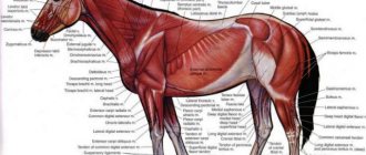
Muscles and skin
The muscle system ensures the dog's mobility. There are 3 types of muscles in the body:
- smooth – located on the walls of all blood vessels;
- striated – attached to the skeleton; dogs have more than 180 single and group muscles of this type;
- cardiac.
All muscle fibers are penetrated by a huge number of nerve endings that transmit impulses to the brain. When contracting, heat is released, which is why in hot weather during activity the body overheats.
Skin provides many functions in a dog's body. It plays the role of a natural sensor that reacts to any external changes, and also protects muscles from injury and regulates body temperature. All layers of the dermis are penetrated by blood and lymphatic vessels, sebaceous and hepatoid glands, and on the surface layer - the epidermis - there are hair roots. The type of coat, its color and structure are determined by breed standards.
Photo: wikimedia.org
Main organs and systems
The structure of a dog’s internal organs is designed in such a way that their body has several important organs and systems, which are listed below:
- Internal organ systems responsible for respiration, excretion, reproduction and digestion.
- Lymph and circulatory systems.
- The apparatus responsible for moving the body, which consists of muscles, bones and ligaments.
- The immune system.
- Skin covering.
- System of endocrine glands.
- Sense organs.
- Nervous system.
Management of the entire body as a whole
To control all body systems, the nervous, circulatory, immune, lymphatic, hormonal, skin and sensory systems are used.
The system is divided into central and vegetative. It consists of nerve fibers. Thanks to its high development, dogs have enhanced senses such as smell, vision and hearing. The central nervous system, together with the cerebral cortex, through congenital and acquired reflexes during life, regulates all systems of the dog’s body.
Blood
The cardiovascular system includes the heart and blood vessels: arterial, coming from the heart, and venous, coming to this organ. The main arterial vessel is called the aorta. The cardiovascular system is designed to supply all organs and cells of the body with oxygen and nutrients and remove waste products of metabolism. The location of the heart is the chest. It is located on the left side of it.
Sense organs and skin
External and internal influences are perceived and analyzed by the senses. A dog has five senses: visual, auditory, olfactory, gustatory and tactile. The optic eye consists of the eye with the pupil, eye muscles and nerves.
The auditory analyzer includes an ear, the structure of which is such that it not only perceives vibrations of sound waves, turning them into sound, but also has the function of correct orientation in space - balance. The sense of smell in dogs is very developed, its acuteness depends on individual characteristics and training. Taste buds are located on the dog's tongue and serve to analyze the composition and quality of substances that enter the mouth.
The cutaneous organ of touch represents primarily a barrier between the external environment and the internal system of the dog’s body. The tactile function protects the organs from adverse effects. Skin composition:
- Subcutaneous tissue.
- Epidermis.
- Wool is a derivative of leather.
Knowledge of the anatomy of the canine body allows us to better understand the reasons that prompt our pets to behave in one way or another.
Dog bones
The Dog Anatomy Atlas includes the skeleton of this animal. It consists of 29 types of bones. All of them are listed below:
- Upper jaw.
- Lower jaw.
- Thoracic vertebrae.
- Skull box.
- Metatarsus.
- Parietal bone.
- Cervical vertebrae.
- Lumbar vertebrae.
- Occipital protuberance.
- Tail vertebrae.
- Brachial bone.
- Bones of the forearm.
- Carpal bones.
- Pastern.
- Phalanges of the fingers.
- Shoulder blades.
- Ribs.
- Hip joint.
- Femur.
- Knee-joint.
- Sternum.
- Rib cartilage.
- Tibia.
- Hock joint.
- Tarsus.
- Fingers.
- Tibia.
- Hip bone.
- Heel bone.
Features of the anatomy and physiology of dogs
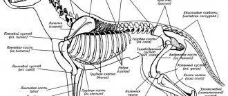
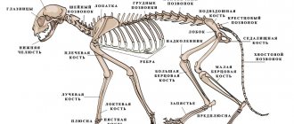
Skeleton of a dog The skeleton plays an important role in the life of the body. It serves as a lever of movement, support for soft parts of the body, protection, a place for the development of hematopoietic organs, and also participates in metabolic and biochemical processes in the body. The skeleton of a dog is unique in its structure. Distinctive features of the skeletal system are strength and lightness compared to other tissues. Young animals have more elastic bones than older ones. As we age, bones become more brittle. The skeleton consists of 247 bones and 262 joints.
The skull consists of the facial and cranial bones. The lower jaw is attached to the skull by a joint, driven by powerful masticatory muscles. The upper and lower jaws contain teeth. An adult dog has 42 teeth, while puppies have 28 (up to 32) baby teeth. Sometimes in a set of teeth there are fewer teeth developed than 42 (oligodontia), sometimes there are more teeth (hyperdontia).
The thoracic limb begins with the scapula, then the humerus, forearm, wrist (7 carpal bones), metacarpus (5 metacarpal bones). The fingers are equipped with strong, non-retractable claws at the end.
The pelvic (hind) limb begins with the femur, passes into the tibia (tibia and fibula), then into the tarsus (consists of 7 bones). This is followed by the metatarsus (of 4-5 metatarsal bones), then 4 phalangeal toes, ending in claws. Sometimes a rudimentary (dewclaw) finger grows from the inside. It is usually amputated at a young age. The pelvic limb has an articular connection with the pelvis and is fixed by the muscles of the hip group.
The structure of the dog's body
Many dog breeders are interested in the question: what is the topography of internal organs in dogs? A dog is a mammal. Their skeleton has a typical structure and the same sections.
Mammals have 7 cervical vertebrae. Giraffes, which have very long necks, and whales, which have no necks at all, have the same number of vertebrae as dogs.
The thoracic vertebrae (depending on the breed of dog, their number varies from 12 to 15 pieces) together with the ribs and sternum are part of the chest.
The lumbar spine, which is responsible for protecting and fixing the dog’s internal organs (photo can be seen in this article), consists of six movable vertebrae. The sacral spine consists of 4 vertebrae connected to the pelvis.
Depending on the breed of the dog, the number of vertebrae in the tail of the skeleton can vary from three to several dozen. The length of the tail depends on the number of bones.
The skeleton of the forelimbs consists of two shoulder blades, which are connected by crow bones and two clavicles.
The supporting part of the hind limbs consists of the pelvic bone, which was formed from three fused bones. It is noteworthy that many mammals, including dogs, have well-developed muscles of the pelvis and limbs.
Video “How do dogs see the world with their nose?”
We have already mentioned how much information our four-legged friends receive through their noses. But this video, which completes your introduction to canine anatomy, will tell you something more interesting about the hypersensitive dog nose!
Sorry, there are no surveys available at this time.
Was this article helpful?
Thank you for your opinion!
The article was useful. Please share the information with your friends.
Yes
No
X
Please write what is wrong and leave recommendations on the article
Cancel reply
Rate the benefit of the article: Rate the author ( 3 votes, average: 3.67 out of 5)
Discuss the article:
Dog's oral cavity
The anatomy of dogs (the photos in the article confirm this) is designed in such a way that they have teeth and a tongue in the oral cavity. The latter serves to determine the taste of food. Its surface is covered with papillae, at the end of which taste nerves are located. The tongue is able to move food in the mouth to moisten it with saliva and chew it.
The anatomy of the internal organs of dogs is designed in such a way that their teeth have long roots, which are placed in the sockets of the jaw. Each tooth has a complex structure and consists of dentin. The outside is covered with enamel. The sharpest teeth (incisors) are located in the front, and powerful fangs are located on the sides of the mouth. The molars are located at the back of the mouth.
In order to securely hold prey and thoroughly chew meat and grind bones, the dog has very well developed muscles responsible for the movement of the lower jaw.
The anatomy of the internal organs of dogs is designed in such a way that the puppies of this animal first grow milk teeth, which fall out after a few months. Subsequently, permanent ones grow to replace them.
All of a dog's teeth are needed to perform specific tasks. So, indigenous ones are used to tear large pieces of meat.
Molars located along the edges have blunt tips. They help grind solid plant foods. Sharply sharpened incisors are designed to separate meat from bones.
Internal organs
The anatomy of a dog’s internal organs is arranged in such a way that in the body of this animal they are represented by 18 main ones. All of them are listed below:
- Oral cavity.
- Anal glands.
- Genital organs.
- Anal hole.
- Heart.
- Nasal cavity.
- Large intestine.
- Kidneys.
- Small intestine.
- Lungs.
- Cerebellum.
- Spinal cord.
- Brain.
- Liver.
- Bladder.
- Trachea.
- Spleen.
- Esophagus.
The anatomy of a dog's internal organs is designed in such a way that their stomach consists of one chamber for processing food. The intestine consists of three types of intestines: rectum, large and small. Once in the intestines, food dissolves under the influence of the secretions of the digestive glands, as well as juices that come from the pancreas and liver.
The anatomy of a dog's internal organs is designed in such a way that the dog's chest cavity is separated by a muscular septum from the abdominal cavity. When the intercostal muscles contract, the chest enlarges. The fins move forward while gaining air. The diaphragm becomes flat. When exhalation occurs, the ribs lower. The chest narrows, which forces air out of the dog's lungs. Exhalation occurs.
The anatomical structure of a dog’s internal organs is such that, regardless of breed, they have a four-chambered heart. It consists of two atria and two ventricles. Blood moves through the systemic and pulmonary circulation.
Due to the anatomical structure of the dog’s internal organs, urine is released when the kidneys work. This paired organ is located on the sides of the lumbar vertebrae. The produced urine enters the bladder, and from there through the canal it is periodically discharged out in small portions.
The successful arrangement of the dog's internal organs allows for rapid metabolism. Body temperature in healthy dogs should be the same. In puppies it is about 38.5 degrees. In adult dogs, the normal body temperature is approximately 37.5 degrees.
The brain is also included in the anatomical structure of the dog’s internal organs. It is located inside the skull and consists of two hemispheres. The hemispheres of the brain have a layer of nerve cells that form its cortex. It is enlarged and contains convolutions. The more there are, the better developed the animal’s brain activity. This department controls complex body movements.
The structure of a dog’s internal organs (you can see photos with inscriptions of all organs in the article) allows these animals to sense five senses:
- touch;
- hearing;
- taste;
- sense of smell;
- vision.
Due to the structural features of the internal organs of dogs, they have a well-developed sense of smell, which helps them search for their prey or a relative even at a great distance. Most dogs' hearing is also well developed. The ears pick up even the quietest sounds.
Long hairs (vibrissae) that grow near the nose and eyes are responsible for the sense of touch.
When in contact with any object or prey, the dog uses three of its senses at once: touch, sight and smell. They need to constantly be in contact with the outside world. They remember their experiences and hone their skills.
Changes in nature contribute to changes in behavior in animals, they develop new reflexes. This ability allows this species of mammal to adapt to various conditions and helps them survive in the surrounding world.
Early dog play (chasing, running and wrestling) provides excellent training for honing individual defense and attack skills.
How does the dog perceive the world around him?
The senses of four-legged pets are more developed than those of humans. Their sense of smell is especially valuable. For this reason, dogs often work as bloodhounds at customs and in the police.
Grandma, why are your eyes so big?
The structure of a dog's visual organ is similar to that of a human. It also includes the cornea, lens and retina. Only the macula, which is responsible for the acuity of central vision, is missing. Because of this, animals have difficulty seeing an object located at a distance of more than 25 cm. Despite this, they are excellent at oriented in the dark and catch moving objects at a distance of 1.6 km. That is, the dog may not notice its owner standing too far away, but can easily distinguish the movement of his hand.
The world seen by four-legged pets is not black and white. For them, only 2 colors are not distinguishable: red and green.
Grandma, why do you have big ears?
The sensitivity of a dog's hearing is much higher than that of a human. Animals detect ultrasound, but do not tolerate its long-term exposure very well. For this reason, these sound waves are used for repelling or training.
Sound enters the outer part of the ear, is processed in the middle and is transmitted through the inner ear directly to the brain. At night, the hearing sensitivity of four-legged pets increases. If during the day they hear at a distance of up to 50 m, then at night - at a distance of up to 150 m. Because of this, dogs living on the street often bark at sounds that irritate them after dark.
The nose is the main sense of smell
A dog's nose can distinguish up to 1 million odors at a distance of up to 1 km. This is due to the huge number of receptors, amounting to about 125 million. For comparison, a person has 25 times less of them.
Tongue and taste
Four-legged pets are second only to us in the ability to sense taste. They will not become gourmets, since their taste buds number only 1,700, while humans have as many as 9,000.
Dogs have a particular dislike for bitter and sour tastes. Although, if there is a tasty smell, they can swallow the hated product, trying not to chew it.
How to measure a dog's temperature
Before measuring your pet's body temperature, shake the thermometer and then lubricate its tip with baby cream or Vaseline. Then it needs to be inserted 2 cm into the rectum, after first lifting the animal’s tail.
The thermometer must be held and not allowed to sit down. The thermometer can be pulled out after 5 minutes. After each measurement, the medical measuring device should be disinfected.
Breath measurement
The respiratory rate can be determined by correctly counting the number of inhalations and exhalations of the animal within one minute. For an accurate calculation, place your hand on the dog's chest or follow the movement of its nose wings. A healthy dog takes ten to twenty breaths in and out for one minute at rest.
The anatomy of a dog’s internal organs is designed in such a way that breathing becomes faster if the animal is running. Also, the number of breaths increases if the dog is scared or excited about something. The breathing rate may change depending on the time of day and weather changes. Due to the structural features of a dog’s internal organs, in the heat, its breathing becomes significantly faster.
Puppies at an early age breathe much more frequently than adults.
Pulse counting
The anatomy of the structure of dogs is designed in such a way that the frequency of their heartbeat is most easily calculated if you place your palm on the animal’s chest.
The pulse can be felt by placing your fingers on the femoral artery. It is located on the inner thigh. The heart of a healthy animal at rest for half an hour should contract at a rate of 60 to 120 beats per minute.
The heart works much faster if the dog is exposed to physical activity, is excited, or feels pain. Also, the heart muscle contracts more often if the animal has an elevated temperature.
Small puppies' hearts work faster than adult dogs'.
Thoracic region
The thoracic section of the body includes a fairly convex rib cage formed by the thoracic vertebrae, 13 pairs of ribs and the breastbone (sternum). The deep chest (measured from the shoulder to the last rib) makes up 2/3 of the dog's total body length. The sternum forms an arc of large radius. It forms the space in which the heart and lungs are located and should be wide enough for dogs undergoing severe physical stress.
Medicines
To provide first aid when identifying symptoms of internal organ damage in a dog, each owner needs to have a veterinary first aid kit at home. Do not forget to check the expiration dates of the medications contained in it. Expired medications must be promptly replaced with new ones. First of all, such a first aid kit should contain:
- Activated carbon.
- Sterile cotton wool.
- Thermometer.
- Iodine.
- Syringes.
- Band-Aid.
- Antipyretic.
- Bandages.
- Syringe.
- Analgin.
Use of drugs
Every dog owner needs to know how to properly give their pet the necessary medications to treat ailments. Some dog owners face difficulties when their dog does not want to take his medication. As a rule, adding drugs to food does not produce any results. The pet quickly learns to take away the tasty morsels, leaving the tablets uneaten. In order to facilitate the procedure for taking tablets and mixtures into the animal’s body, you should resort to some tricky techniques:
- Use a syringe. In this case, it is necessary to pour the medicine into the mouth at the same time as the owner squeezes the dog’s jaws. This will prevent the animal from closing its jaws.
- Once the tablet or powder is on the tip of the tongue, the dog needs to hold its mouth tightly with your hands to prevent the medicine from falling back out. The sick animal must be kept in this way for several minutes until the product enters the esophagus.
- Powdery substances should be poured onto the dog’s tongue, and after a while, give him a drink of clean water.
- To force the dog to drink the mixture, you should throw the animal's head up, and then try to pour the liquid behind the jaw. Now all that remains is to wait until the pet reflexively swallows the medicinal product.
- Applying the products externally directly to the dog’s body does not cause much trouble. However, after applying the cream or gel, you must put a protective collar on the animal. This device will prevent the dog from licking the medicine from the body.
- Rectal and vaginal administration of drugs is carried out using suppositories and special enemas for animals. After successfully introducing the drug into the colon, it is necessary to press the tail against the anus for a while.
- Instilling drugs into the eyes is a complex procedure. It is advisable to do it together: one holds the dog’s muzzle, and the second should apply the medicine, while holding the dog’s eyelids open with one hand.
- If you need to inject a syringe with the drug under the skin of an animal, you need to buy a syringe with a thin and short needle at the pharmacy. For the medicine to work, you need to gather a fold near the withers with two fingers, and then insert the needle to a depth of 2 cm. Then you need to inject the liquid by slowly pressing the syringe plunger.
- Sometimes pet owners have to give intravenous injections to their pets themselves. For this procedure, it is necessary to find the small saphenous vein in the dog's lower leg. A tourniquet must be applied above the puncture site, tightly tightening the paw. Only after this can the vein be pierced with a needle. After this, the drug can be administered. It is very important to hold the animal’s paw during this procedure, otherwise it may be injured.
All other procedures, such as installation of IVs, intracavitary injections, injection of the drug into the heart muscle, blood transfusion, ultrasound of internal organs in dogs should only be performed by a qualified veterinarian.
general characteristics
To make it easier to understand the anatomy of a dog, the dog’s body parts are conventionally divided into four main components, namely:
- head. This part includes the brain and facial zones, as well as the sensory organs;
- neck. There are upper and lower regions;
- torso. This category is more expanded and is considered from the withers to the tail;
- limbs. This category includes the front and hind legs.
Unfolded skeleton of a dog
The external characteristics of the dog, its body parts and their location, which are inherent in the breed and gender, are called the exterior and look like the signature of the breed.
Note! In addition to the general idea, it would not hurt for the dog owner to study the textbook, authored by Volmerhaus and Frewein, which provides a diagram and location of all the animal’s organs. The practical textbook is regarded as a guide for dog breeders.