Anatomy of the hearing organ Etiology (causes of occurrence) of otitis Symptoms of external otitis Diagnosis Treatment Cleaning the ears of dogs and cats Otitis in a dog or cat (Greek otos - ear; itis - inflammation) is a group of diseases that combines inflammatory diseases of the ear of various etiologies.
Depending on the affected department there are:
- - otitis externa
- otitis media
- internal otitis media
Otitis externa
- inflammation of the external auditory canal - belongs to the group of dermatological diseases, since the initial part of the auditory canal is lined with skin. According to statistics, up to 50% of dermatologist patients have otitis media, either as a primary problem or as a secondary problem (a consequence of a primary disease, for example, atopy).
Despite the fact that the diagnosis of otitis media is often made based on examination, clarifying the cause of its occurrence and, accordingly, choosing specific therapy may require additional diagnostics. Often, otitis media, especially in an advanced stage, are quite difficult to treat.
When diagnosing and treating otitis in a dog, the approach to the patient should be as thorough as the approach to any other patient with systemic dermatological pathology; the examination should not be limited directly to the ear area, but should affect the entire skin of the animal.
Anatomy of the hearing organ
The ear, as an organ of hearing, is conventionally divided into three sections: external, middle (represents a sound-conducting apparatus) and internal (sound-receiving apparatus). The outer ear includes the movable pinna and the external auditory canal, which is a derivative of the skin. The skin of the auditory canal contains a large number of ceruminous glands, which secrete a secretion - earwax, which plays an important protective function.
The external auditory canal in a dog is shaped like the letter L, and consists of a vertical and horizontal part, invisible during normal examination. The fact that the external auditory canal is narrow, long enough and has a bend protects the eardrum from damage and provides mechanical protection to the deeper structures of the ear from external influences. The eardrum acts as a sound absorber and separates the outer ear from the middle ear.
The middle ear is a tympanic cavity that contains three auditory ossicles: the malleus, the incus and the stapes. The middle ear cavity communicates with the nasopharynx. The main function of the middle ear is to conduct sounds from the eardrum through the auditory ossicles to the oval window, which leads to the vestibule of the labyrinth of the inner ear.
The inner ear is located in the temporal part of the parietal bone and consists of the bony and membranous labyrinths, within which the organs of hearing (cochlea) and balance (vestibular apparatus) are located.
Some anatomical features of the structure of the external auditory canal in dogs can contribute to the development of otitis: hanging ears, complicating the ventilation of the auditory canal; a curved ear canal, making it difficult for water to get out of the ear; breed predisposition (in some dog breeds, such as Labradors and Cockers, the skin of the external auditory canal contains a large number of glands).
Here are some of the causes of ear problems in dogs:
- Ear mite. This is a tiny mite that lives and reproduces in the ear canal and causes itching and an allergic reaction in the dog.
- Foreign bodies. This definition includes everything that ends up in the ear, although it should not be there. In this case, very strong irritation and discomfort are characteristic.
- Eczema. This is the name for skin inflammation caused by a number of reasons.
- Tumor. Sometimes a skin tumor develops in the ear canal, blocking the canal and interfering with the normal functioning of the ear.
Etiology (causes of occurrence) of otitis media
Primary reasons:
- allergy (hypersensitivity, atopy)
- parasitic diseases (otodectosis, notoedrosis, cheyletiellosis, demodicosis)
- autoimmune processes and diseases (pemphigus foliaceus, discoid lupus erythematosus)
- some viral diseases (otitis media associated with canine distemper virus has been described)
- foreign objects (especially small cereal seeds)
- neoplasms (eg, ceruminous gland tumors)
- endocrinopathies
Secondary causes:
- bacterial inflammation
- fungal diseases (Malassezia dermatitis, candidiasis)
Predisposing factors:
- the structure of the ear canal (drooping ears, congenital stenoses (Shar Pei), an abundance of hair in the external auditory canal), causing impaired ventilation of the ear canal
- high humidity, warm season
- swimming, diving, especially in polluted waters
mechanical damage to the external auditory canal (due to improper ear cleaning)
Otodectosis
Parasitic disease. Caused by the microscopic mite Otodectes. Dogs, cats and rabbits get sick. Characterized by severe itching, the dog scratches its ears with its paws and rubs against various objects. Mites cause inflammation, irritation and damage to the delicate skin of the ear canal, which contributes to increased secretions and the proliferation of fungi and microbes. As the inflammatory process develops, first serous and then purulent-ichorous exudate is released from the ear, which dries out in the form of crusts and scabs, impeding free outflow. The waste products of mites color the discharge brown and black. The discharge has a peculiar unpleasant odor.
The diagnosis of “otodectosis” is made on the basis of clinical data and laboratory tests. Microscopy of ear scrapings is available in any clinic that has a light microscope.
Otodetis mites are sensitive to most acaricides, so they can be easily destroyed with proper treatment. In order for anti-mite drugs to penetrate the parasites, it is necessary to clear the ear canal of crusts, wax and exudate. Otherwise, the medicine will simply dissolve in the contents of the ear and will not have any effect.
Otodectosis can be easily cured only at the very beginning. In the process of development, otitis media is added to the parasitic disease, which is much more difficult to cure.
Diagnosis of otitis media
Basic diagnostic methods
- inspection
- otoscopy (visual examination of the vertical auditory canal using a special device - an otoscope)
- cytological examination is an examination of a specially stained smear from the external auditory canal, which allows one to assess which cells and in what quantity are present in the exudate, as well as identify parasites, bacteria, and opportunistic fungi. This study may be required every 2-3 weeks to monitor treatment and response to therapy.
Additional diagnostic methods
- bacteriological examination (culture, determines the sensitivity of flora to antibiotics)
- endoscopic examination of the deep structures of the auditory canal (horizontal part) and eardrum (myringoscopy) - the study is carried out under general anesthesia.
- radiography of the tympanic cavity - as a rule, for a high-quality image, complete immobility of the animal and symmetrical positioning are necessary, which is only possible under sedation.
- MRI (under general anesthesia).
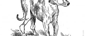
Skeleton or skeletal system
The skeleton is the frame that holds all the internal organs, as well as the muscles of the dog. The structure of a dog’s skeleton can be easily represented in the diagram as two lines:
- axial, which includes 109 bones (skull and spine with ribs);
- peripheral, consisting of 180 limb bones.
Over the course of an animal's life, the composition of bone tissue changes. Therefore, in puppies and teenage dogs, the bones are more elastic and lighter, but in old age there is a high probability of increasing the risk of fragility and loss of strength. The condition of the bones and teeth is used to judge the general health of the dog.
Structure of the skull
The facial and cerebral parts of the skull are distinguished; both include paired and unpaired bones. In total, the skull consists of 27 bones connected to each other by cartilage tissue. With age, the cartilage becomes ossified, and mobility is maintained only in the lower jaw area so that the dog can chew food.
The figure shows both paired and unpaired bones of the skull.
Based on the type of skull, dogs are divided into representatives of dolichocephalic (prominent representatives are Italian Greyhounds and Greyhounds) and brachycephalic breeds (for example, pugs, dwarf Spitz). The biggest differences between them are noticeable in the structure of the facial part of the skull. So, brachycephals have a flattened muzzle and a protruding jaw. It is these characteristics that have been specially cultivated by breeders over the years to make the breeds recognizable. But such features are associated with certain health problems for pets.
Structure of teeth
Teeth are not only an important part of a dog's appearance. First of all, teeth are necessary for biting and grinding food, protecting the owner and, if necessary, attacking the enemy.
Puppies are born without teeth. At the age of two to three weeks, the first baby teeth emerge through the gums. Closer to 4-5 months, they begin to fall out to make room for permanent ones. Instead of 28 milk teeth, by the age of one and a half years, the jaw should have 42 permanent teeth. Deviations from the schedule are often due to unbalanced nutrition or breed characteristics.
The dental formula of an adult dog includes 42 teeth, with 20 located on top and 22 on the bottom.
The permanent set of teeth for an adult dog includes:
- Incisors – 6 on each jaw.
- Fangs – 2 on top and bottom. They are dangerous weapons in fights.
- There are 4 premolars on both branches of the jaws.
- Two molars on each branch of the upper jaw, as well as three below, for a total of 10 pieces.
Dental arcade of a dog
The tooth consists of a crown, neck and root. The crown protrudes significantly above the gum; each type of tooth has its own shape. Dentin is the main dental tissue; in the crown area it is covered with enamel, and in the root zone dentin is covered with cement. Inside the tooth there is a cavity divided into the coronal space and the root canal itself.
The number of teeth, their condition and bite (or occlusion) directly affect the health of the dog. The following typology of bites is distinguished:
- Scissor-shaped.
- Pincer-shaped.
- Snack.
- Underbite.
The most common type is the first type of bite.
Spine structure
The spinal column is the axis of the skeleton. A skull is attached to it on one side, and it ends in a tail. Also on the sides, ribs and limbs are attached to it using cartilage tissue.
The structure of the spine can be represented as follows:
- Cervical spine - consists of 7 vertebrae, the first two of which (atlas and epistrophy) are especially mobile. They are responsible for head movements.
- The thoracic region includes 13 vertebrae. The ribs are attached to them, forming the chest. Dogs have 9 pairs of real and 4 pairs of false ribs.
- The lumbar region also consists of 7 vertebrae.
- The sacrum is a sacral bone fused from 3 vertebrae.
The dog's tail, which is a logical continuation of the spine, consists of 20-23 vertebrae. The most developed and mobile are the first five. Previously, representatives of certain breeds had their tails docked, but now such actions are not supported by the global canine community.
Separately, we should consider the structure of the dog’s penis, since it also includes a bone - the baculum, based on the connective tissue of the penis. The baculum is located at the front of the penis. Its upper edge is convex, and below there is a groove with a passing urogenital canal. In dogs, the penis belongs to both the reproductive system and the excretory system, since the urinary canal is also the vas deferens.
Limb structure
The limbs of dogs are characterized by the complexity of their structure. The front legs are an extension of the scapula, which is attached to the spine by developed shoulder muscles. The shoulder blade goes into the humerus, then the forearm and wrist joint. The forearm consists of the radius and ulna bones, and the metacarpus includes 5 bones. The hind legs are formed by the thigh, stifle, shin, hock, metatarsus and paw.
The structure of the paws can also be represented in this way:
- Pads that act as shock absorbers. They reduce stress on bones and joints and also help maintain balance. The pads consist of an impressive layer of fatty tissue, so dogs do not freeze in cold seasons, and heat is well retained in their paws.
- Pets' fingers have different numbers of phalanges. 4 fingers are three phalanges, and one is only two. An animal cannot move them in the same way as a person due to the limited space between the fingers. Normally, dogs have 5 toes on their front paws, and 4 on their hind paws. There are also rudimentary toes - dewclaws, located on the hind paws just above the foot. They do not carry any functional load, but in some cases they can be a sign of a high quality representative of the breed. This is true for Briards, Beaucerons or Pyrenean Mastiffs.
- Dog claws, unlike cat claws, do not retract into the pads and consist of hard, keratinized tissue and pulp, an area with a significant number of blood vessels and nerve endings. It is important to be very careful when trimming nails so as not to cause harm or pain to the dog. It is also necessary to monitor the condition and length of the claws, since there is a direct connection between them and the musculoskeletal system. Long claws do not allow a four-legged pet to walk normally; because of them, the skeleton can even begin to deform.
Treatment of otitis media
Treatment of otitis depends on the causes that caused it (both primary and secondary).
1. For parasitic otitis, antiparasitic drops are used on the withers 2 times with an interval of 3 weeks and daily sanitation of the ear canal is carried out. When parasitic otitis is complicated by a secondary bacterial or fungal infection, specific local (in advanced cases - systemic) therapy is carried out.
2. For allergic and other otitis, the root cause is eliminated, and local (in severe cases systemic) antibiotics or antimycotics are used together with local anti-inflammatory therapy and daily sanitation of the ear canal.
3. Treatment of verrucous (warty) otitis, which develops with long-term otitis, comes down to the use of conservative and surgical methods.
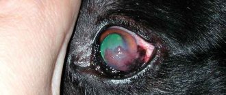
Signs of ear diseases
A caring owner can determine the presence of an ear disease at home by the following signs:
- the dog scratches his ears until they bleed;
- the tips and passages of the ears are damaged and swollen;
- specific discharge from the ear appears with a characteristic unpleasant odor;
- while walking, the animal shakes its head or tilts it to one side;
- touching the ears causes discomfort or pain to the pet.
In addition, the painful condition of the hearing organs affects the dog’s activity. If the owner notices a change in the pet’s mood, poor physical activity, or refusal to communicate, it’s time to see a veterinarian.
Cleaning the ears of dogs and cats
Ear cleaning is carried out only when indicated, either with warm saline solution or special lotions. A veterinarian should also prescribe the use of lotions, as there may be contraindications for their use.
Cotton swabs should not be used at any stage of ear cleaning. Cotton microparticles lead to additional irritation of the skin of the auditory canal and increased secretion; in addition, the pathological contents of the ear canal are only “compacted” deeper into the horizontal, visually inaccessible part of the canal, aggravating the inflammation.
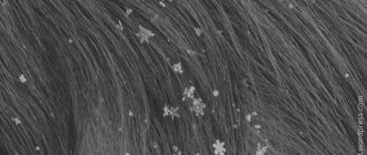
A healthy animal without visible discharge does not need to have its ears cleaned “for prevention.” The norm is a scanty fatty yellowish-gray discharge (“wax”), making the inner surface of the external auditory canal slightly shiny.
Preventative cleaning
may be useful for dogs with stenosis of the auditory canal or poorly ventilated canal (loose-eared, sharpei, etc.), but it should
only be carried out on the recommendation of a doctor
.
If the animal has pathological secretion or an established diagnosis of otitis, it is necessary to pour a small amount of warm (37-38.C) sterile saline solution or a special lotion for cleaning the ears into the ear, keep the animal from immediately shaking its head, lightly massage the ear base, and after a minute allow the dog to shake its head vigorously so that the liquefied ear secretion “flies out” under the influence of centrifugal force. The procedure must be repeated until the solution is clean, after which it is necessary to blot the outside of the ear with a soft, lint-free cloth. You cannot go deeper into the external auditory canal than an adult’s finger can penetrate using cotton swabs and other devices. (c) Veterinary center for the treatment and rehabilitation of animals “Zoostatus”. Varshavskoe highway, 125 building 1. tel. 8 (499) 372-27-37
Prevention of ear diseases
Examine your dog's ears after walks in the park, field, or forest. If you find ticks, plant seeds, or dirt, remove them immediately. In winter, pay attention to the temperature of your ears - their tips become frostbitten very easily. Make sure that when washing, water does not get into the ear canal. Do not allow the animal to dry in a draft after bathing. Do not give your dog sweets, smoked foods and other prohibited foods - they provoke the secretion of earwax. Strictly control the diet of an allergic dog. Clean your pet's ears regularly using mild cleansers, such as Bars lotion. Do not use ear swabs, only cotton or gauze pads. Contact your veterinarian promptly if you have any suspicions.