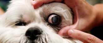
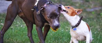
Dogs, like other animals, often suffer from eye diseases. By the condition of the eyes you can always determine whether your dog is healthy or not; the eyes are a “mirror” not only of the soul, but also of the health of the animal. In medicine, the eyes are used to diagnose existing diseases in a person. In medicine, as one of the auxiliary diagnostic methods, there is iridodiagnosis - the diagnosis of diseases in a person using the iris of the eyes. When conducting iridology, special equipment and computer programs are used. When making a diagnosis, doctors take into account changes in the structural state, shape of the color areas of the eye, as well as the mobility of the iris.
Before talking about eye disease and its auxiliary organs, it is necessary to have a general understanding of its structure.
A dog's eyes are located in the orbits - bony sockets formed by the bones of the skull, where they are held by several muscles that ensure their mobility and orientation in different directions.
The dog's eye itself is protected by auxiliary organs - eyelids and glands. The dog has three eyelids. The upper and lower eyelids are folds of skin, the inner surface of the eyelids is lined with mucous membrane. The outside of the eyelids is bordered by eyelashes, which protect the eyes from dust and other foreign particles. A dog's third eyelid is a simple film in the inner corner of the eye that dog owners usually cannot see. This film covers the eye when it is closed or irritated, as well as during nervous disorders.
The eye in the cornea area comes into contact with the external dry environment, so it needs protection of the lacrimal glands, which produce tear fluid - a secret that moisturizes the surface of the cornea. A dog's tears accumulate in the space between the eyelids and the eye and are then drained through a narrow channel that begins at the inner corner of the eye and opens into the nasal cavity. When there is excessive lacrimation or blockage of the tear duct, tears flow from the eyes and, when oxidized, form red stripes on the fur that look like blood.
The eye consists of two parts.
- The anterior portion includes the cornea, iris, and lens. They absorb beams of light from the dog, like a camera lens. The cornea and lens are clear and act like optical lenses, and the iris acts as a diaphragm, regulating the amount of light entering the eye through the pupil (the hole in the iris).
- The back of the eye consists of the vitreous body, the choroid (choroid) and the retina, which converts optical light signals into nerve impulses that are transmitted to the visual center of the brain.
Thinking of the eye as an analogy to a camera, the back of the eye is like a photographic film on which the dog's brain captures the image.
Experts, depending on the cause, divide all eye diseases in dogs into 3 types:
- Infectious – occurring in dogs due to the presence of viral or bacterial diseases, most often as a complication of the underlying disease.
- Non-infectious - due to certain mechanical damage, inflammation as a result of improper eyelash growth, neoplasms, eversion of the eyelids.
- Congenital – include eversion, entropion of the eyelids, deformations of the eyes and lens. Congenital ones are most often found in some dog breeds (Shar Peis).
Distichnaz
With this disease, single or multiple hairs appear in a row on the free edge of the eyelid, which should be hairless.
These hairs appear in a dog only at the 4-6th month of life and can be either very delicate or quite hard. With this disease, several hairs most often grow from one point. This disease is most often recorded in English and American cocker spaniels, boxers, Tibetan terriers, collies, and Pekingese.
Clinical picture. During a clinical examination of a dog, a veterinarian notes profuse lacrimation, constant blinking, blepharospasm, irritating hairs have contact with the cornea of the eye. If a dog has curled eyelashes, keratitis is diagnosed.
The diagnosis of the disease is made based on the above symptoms.
Differential diagnosis. Distichnasis is differentiated from trichiasis, entropion and eversion of the eyelids, allergic conjunctivitis, and keratoconjunctivitis sicca.
Treatment. It is carried out in veterinary clinics by electrolysis under an operating microscope. Excision of the third eyelid.
Trichiasis
Trichiasis is a condition when hair from a dog's eyelids or muzzle gets into the eye, coming into contact with the conjunctiva and cornea. Trichiasis can be primary or secondary. Primary occurs in dogs with medial inversion of the eyelids and a large nasolabial fold. Trichiasis occurs in the following dog breeds: Pekingese, Pugs, English Bulldogs, English Cocker Spaniels, Chow Chows, Shar-Peis.
Clinical picture. During a clinical examination of a dog, a veterinarian notes lacrimation, hairs in contact with the cornea cause blinking in dogs, constant discharge from the eyes, symptoms of keratoconjunctivitis, inflammation of the skin in the area of the nasolabial fold.
The diagnosis is made based on the detection of hair in contact with the cornea, provided there is no other eye pathology.
Differential diagnosis. Trichiasis is differentiated from keratoconjunctivitis sicca, entropion and eversion of the eyelids, dystrichiasis, and ectopic eyelashes.
Treatment. Treatment of the disease is surgical. Temporary improvement can be achieved by trimming the hair that gets into the eye.
Entropion of the eyelids
Entropion is a pathology of the eye in which part of the organ turns inward towards the eyeball. A dog's eyelid inversion can be either upper or lower, one-sided or two-sided.
Unilateral inversion of the eyelid margin is most often the result of heredity and appears in a dog in the first year of life. Congenital entropion occurs in puppies after the eyes open in some breeds with excessively folded skin on the head (chow chow, shar pei).
In this disease, the eyelashes, hair and skin of the eyelid rub against the surface of the cornea, causing inflammation and irritation.
Clinical picture. During a clinical examination, the veterinarian notes the leakage of liquid secretion from the eye, the dog has photophobia (to an electric light bulb, the sun), the dog rubs its eyes with its paw, blinking, and there may be an eye tic.
Treatment. Treatment of entropion of the eyelids is surgical.
Animals at risk
The risk group includes animals that are most vulnerable to the listed disorders. These include:
- brachycephals with bulging eyes (Japanese chins, Shih Tzus, Pekingese, pugs);
- breeds with folded skin (chow-chow, sharpei, English bulldogs);
- pets with long hair (Yorkshire terriers, Afghan hounds, lapdogs).
In the first case, vulnerability to external factors is explained by the visual organ being too large. In the second - skin folds on the face, increasing the likelihood of inflammatory processes due to accumulating dirt and dust. In the third - long hair, fraught with frequent irritation and injury.
Eversion of the eyelids
With eversion of the eyelids, the edge of the eyelid turns outward, while the mucous membrane (conjunctiva) of the eyelid is exposed.
This pathology occurs in dogs with too large palpebral fissure and excess, easily removable skin in the head area.
Cause. Mechanical eversion of the eyelids in a dog occurs as a result of pathological changes in the eyelid itself, as well as tissue scarring after injuries or surgery.
Paralytic ectropion occurs in dogs as a result of facial paralysis.
Clinical picture. During a clinical examination, the veterinarian notes incomplete closure of the eyelids, discharge from the eyes, and inflammation of the conjunctiva.
Treatment. Treatment for this pathology should be aimed at eliminating the cause that caused and maintains the ectropion of the eyelids (removal of a neoplasm, conjunctivitis, facial paralysis, surgical removal).
When to see a doctor
Eye disease in cats and other animals, general symptoms that should alert the owner:
- poor orientation, especially in an unfamiliar space;
- discharge from the eye: fibrous, purulent;
- dilation of the pupil, clouding of the membranes;
- no reaction to bright sun or light.
Treatment of eye diseases in animals in Moscow
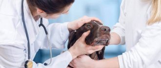
Dr. Vorontsov’s veterinary center employs highly qualified doctors who will conduct a thorough diagnosis of eye diseases. Eye diseases in cats and dogs develop gradually, the main thing is to notice the first symptoms in time and go to an ophthalmologist. Timely contacting a specialist will allow you not only to carry out prevention, but also to develop tactics for adequate treatment of eye diseases of your pet.
Our veterinary clinic is located on Kashirskoye Highway, near the Moscow Ring Road, exact address: State Farm named after. Lenina, building 3a (direction map), metro stations Domodedovskaya, Orekhovo, Zyablikovo, Krasnogvardeyskaya.
- MORE ABOUT TREATMENT OF EYE DISEASES AND PRICES FOR SERVICES
- ALL PRICES AND SERVICES
Sign up by phone:
+7(495) 740-48-59 +7(936) 001-03-04
Blepharitis
Blepharitis is inflammation of the eyelids.
Cause. Unilateral blepharitis in a dog occurs due to injury and local infection. Bilateral blepharitis occurs as a result of allergies, including food allergies in animals, staphylococcal infections in dogs, demodicosis (treatment and prevention of demodicosis in dogs), mycoses and systemic diseases.
Clinical picture. During a clinical examination, the veterinarian notes redness, swelling, itching, scaling, loss of eyelashes and hair, erosion and ulcers in the eyelid area of a sick dog.
Treatment. In the case where the cause of blepharitis is an allergy, dog owners should exclude its contact with the allergen and use antihypertensive drugs (diazolin, suprastin, diphenhydramine, tavegil) in treatment. For staphylococcal infections - antibiotics. For demodicosis, anti-mite drugs.
What to do at home
Many owners have a question: “How to help a dog at home?” The answer is clear: strictly follow the doctor’s instructions and do not self-medicate, as it can lead to irreversible consequences. The maximum that can be done is to create a comfortable environment for the dog, provide it with good care and quality feeding.
As the disease progresses and the dog becomes completely helpless, the owner must be prepared for the fact that he will have to care for the pet, remove feces in a timely manner, and perform anti-bedsore massage.
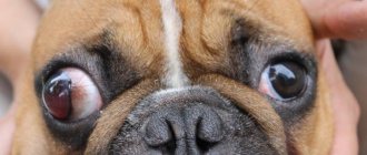
Exophthalmos (protrusion of the eyeball)
Exophthalmos in dogs can be species-specific and is typical for dogs of brachycephalic breeds, with a normal eyeball size, a flat orbit and an overly large palpebral fissure.
Acquired exophthalmos - in which a normal-sized eyeball moves forward due to space-demanding processes in the orbit or its immediate surroundings, or due to an increase in the size of the eyeball as a result of glaucoma in a dog.
Clinical picture. During a clinical examination, the veterinarian notes that the dog has strabismus, an abnormally wide palpebral fissure with protrusion of the eyeball; in some dogs, prolapse of the third eyelid is possible.
Treatment is only surgical.
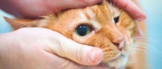
Cataract
The pupil is located in the middle of the eye and is usually clear, but sometimes a cloudy, opaque cataract appears on part or the whole pupil. It blocks light from reaching the back of the eye, resulting in poor vision or blindness, depending on the severity.
Cataracts are often confused with a normal aging change that affects a dog's pupils, called reticular sclerosis. Both conditions give the pupils (usually the black center of the eye) a white, gray, or milky appearance, but a veterinarian can tell the difference with a standard eye exam.
Cataract surgery is available for dogs when their vision is severely impaired. If this is not an option, it is important to recognize that most dogs adapt very well to poor vision. A keen sense of smell, hearing, caring owners or a flock in the wild are very helpful.
Convergent strabismus
Convergent strabismus is a noticeable visual deviation from the normal position and joint movement of both eyes of a dog.
Moreover, with paralytic strabismus, the dog's squinting eye does not repeat the movement of the fixed eye.
Cause. Traumatic eye injuries, hypertrophic processes in the orbit (tumors), damage to the central nervous system.
One of the reasons may be congenital underdevelopment of the periorbital muscles, congenital hydrocephalus.
Treatment. Treatment of convergent strabismus involves treating the underlying disease that led to the strabismus.
Causes of eye diseases
A dog's eyes perform an important function - they convert reflected light into nerve impulses, which the brain uses to form images of the world. To do this, all parts of the eye must be healthy.
However, there are some reasons why vision may suffer:
● Heredity.
● Age.
● Immune system disorders.
● Metabolic abnormalities.
● Infections, parasites.
● Injuries.
Diseases of the structures of the eye, eyelids and surrounding tissue can be accompanied by discomfort and/or loss of vision, which subsequently interferes with your dog's normal life and reduces the quality of his life.
Allergic conjunctivitis
Allergic conjunctivitis in dogs occurs as a result of contact with the mucous membrane of the eye of one or another allergen (contact allergy). The allergen can be pollen from flowering plants, dust, etc.
Allergic conjunctivitis in dogs In recent years, allergies to certain food products (food allergy) have often been recorded.
Clinical picture. During a clinical examination, a veterinarian notes in such a dog redness of the mucous membrane of the eyes, mucous discharge from the palpebral fissure. As a result of itching, the dog rubs its paw on the affected eye.
Treatment. If contact dermatitis occurs, it is necessary to rinse the eye affected by inflammation with saline solution or chamomile decoction.
In case of food allergies, it is necessary to exclude the allergic product from the dog’s diet and transfer the dog to a hypoallergenic diet (buckwheat, rice, beef).
The sick dog is prescribed antihistamines (cetirizine, diazolin, suprastin, diphenhydramine, tavegil), and Diamond Eyes eye drops are instilled into the conjunctival sac.
Diagnostics
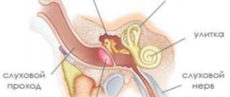
Treatment of vestibular syndrome in dogs begins with diagnosis. To do this, the veterinarian collects anamnesis to find out what factors provoked the development of a disorder of the vestibular apparatus. Additionally, a general and biochemical blood test, magnetic resonance imaging (MRI), X-ray and ultrasound examination of the animal’s internal organs and head are prescribed. Separately, the veterinarian examines the condition of the dog’s middle and inner ear.
On a note! If necessary, a bacteriological examination of the contents of the middle ear is prescribed.
Purulent conjunctivitis
Purulent conjunctivitis in a dog develops due to the entry of various pathogenic microorganisms into the conjunctiva. Purulent conjunctivitis is one of the symptoms of carnivore plague......
Clinical picture. During a clinical examination, a veterinarian notes reddening of the conjunctiva, its swelling, and purulent discharge from the eye of a sick dog.
Treatment. With this form of conjunctivitis, a sick dog is treated with eye drops and ointments that contain antibiotics. Tetracycline eye ointment and Ciprovet drops are widely used. Before applying eye drops and eye ointment, it is necessary to clean the affected eyes of exudate.
Ulcerative keratitis
Severe inflammation of the cornea of the eye. Causes: mechanical damage and trauma (burns, foreign bodies, bruises), infections, reactions to drugs, allergies, vitamin deficiency. It may occur due to diseases of the immune system, in particular diabetes.
Vivid symptoms: suppuration and tearing, fear of light, a cloudy area in the affected area, the presence of ulcers on the cornea, frequent blinking, redness of the white. The consequence of improper treatment is glaucoma, cataracts, blindness.
| List of drugs for treatment | ||
| Name of medicine | Price | Release form and application |
| Levomycytin | 50-60 rubles per bottle | Eye drops are used 4 times a day, 2-3 drops into the conjunctiva; |
| Erythromycin ointment | 90 rubles per tube | Antibiotic ointment is applied around the eyes and on the eyelid of the dog 2-3 times a day, the timing of use depends on the severity of the disease |
Follicular conjunctivitis
This form of conjunctivitis is most typical of chronic conjunctivitis and often develops in a dog when toxic substances get into the eye.
Clinical picture. During a clinical examination, a veterinarian reveals many bubbles with transparent contents on the mucous membrane of the conjunctiva. Mucous discharge comes from the palpebral fissure. The conjunctiva itself is crimson in color, and the dog's inflamed eye is squinted.
Treatment. When treating this form of conjunctivitis, eye ointments containing an antibiotic are used. In severe cases of the disease, specialists are forced to resort to excision of the conjunctiva and subsequent symptomatic treatment.
Panophthalmitis in dogs
This disease is characterized by purulent inflammation of all tissues and membranes of the eye. The cause of the pathology is most often mechanical damage to the eye and the introduction of pathogenic microflora onto the damaged surface. Clinically, with panophthalmitis, there is an increase in body temperature, general depression, loss of appetite, the eye is swollen, clouding of the cornea, discharge of purulent contents, increased pain may be observed, over time the eye atrophies and loss of vision occurs.
As treatment, a broad-spectrum antibiotic is used systemically in the maximum permissible dose, however, this often does not lead to recovery and removal of the affected eye is required.
Disease of the lacrimal apparatus
Keratoconjunctivitis sicca - this disease is characterized by a very small amount of the tear film of the eye as a result of insufficient or absent tear production. This disease is observed in West Highland white terriers and is inherited by their offspring. Keratoconjunctivitis sicca in dogs occurs due to disorders of sex hormones, canine distemper, trauma to the frontal part of the skull, neuropathy of the facial nerve, congenital hypoplasia of the lacrimal glands, and from the use of certain medications.
Clinical picture. Veterinary specialists, when conducting a clinical examination of a sick dog, note frequent blinking, dry crusts at the edges of the eye, itching, the presence of mucopurulent discharge from the eyes, viscous mucus is found in the conjunctival sac, follicular conjunctivitis. Later, as the disease progresses, symptoms of ulceration and unevenness of the corneal surface appear, and swelling of the conjunctiva develops. If there are dry crusts in the area of the nostrils on the affected side, we can talk about the presence of damage to the facial nerve in the sick dog.
Treatment. Treatment for this form of keratoconjunctivitis should be aimed at eliminating the underlying cause of the disease. The area of the conjunctiva and cornea is washed generously every two hours with saline before each application of the drug. The inner corners of the eyes of a sick dog are washed with a solution of chamomile or chlorhexidine, since the lacrimal sac in a sick dog is a reservoir for various microorganisms.
During treatment, eye ointment with an antibiotic is used.
Uveitis
Inflammation of the mucous membranes and irises, from which no dog is immune. The reasons are different: trauma, internal chronic diseases, infection or virus. Symptoms: inflammation of the eye, change in iris color (cloudness), decreased vision, photophobia (the dog hides in dark places), severe pain and half-closed eyelids. Treatment is aimed not only at eliminating pain and inflammation, but also at preserving vision.
| List of drugs for treatment | ||
| Name of medicine | Price | Release form and application |
| Rimadyl (base – carprofen) | 500-650 rubles per plate of tablets (10 pieces) | The product is used to reduce inflammation and relieve pain; The drug comes out in the form of a syrup, the dose is selected only on the recommendation of a veterinarian |
| Knuckles | 80-100 rubles per ampoule | Anti-inflammatory substance (not steroid), available in the form of an injection solution based on diclofenac |
Diseases of the cornea.
Keratitis is a disease of the cornea of the eye. The most common types of keratitis in dogs are:
- Purulent superficial keratitis.
- Vascular keratitis.
- Purulent deep keratitis.
The causes of keratitis in dogs are varied:
- Mechanical injuries.
- Burn damage to the ocular surface.
- Hypovitaminosis state.
- Infectious diseases (canine distemper, parvovirus enteritis in dogs, infectious hepatitis in dogs).
- Invasive eye diseases (dirofilariasis).
- Diseases of the endocrine system (diabetes mellitus).
- Weakening of the immune system.
- Genetic predisposition.
- Allergic reactions.
Clinical picture. During a clinical examination of a sick dog, a veterinarian notes in a sick animal:
- Profuse lacrimation from the affected eye.
- Cloudiness of the eye cornea.
- Photophobia.
- Swelling.
- The sclera and conjunctiva are hyperemic.
- Purulent discharge comes from the eye.
- Gray, yellow and white spots appear in the cornea of the eye.
- Redness of the white of the eyes and mucous membranes.
- The ocular membrane is rough.
- The dog blinks frequently.
- Dark smudges appear in the inner corner of the diseased eye.
- The dog becomes nervous, restless or lethargic and depressed, trying to hide from the light, constantly rubbing its eyes with its paws.
If keratitis in a dog is not treated in a timely manner. As the disease begins to progress, inflamed blood vessels grow into the eye cornea, causing it to become lumpy and thickened.
Consequences of keratitis. Keratitis for a dog is fraught with the development of complications such as the development of glaucoma, cataracts, and corneal perforation. Partial or complete loss of vision.
Treatment of keratitis in a dog depends on the cause of the keratitis, as well as on the factors that provoked its development.
Based on this, the clinic’s veterinary specialist prescribes appropriate treatment for the dog. At the same time, in all forms of keratitis, the sick dog’s lacrimal sacs are washed daily with solutions of furatsilin, rivanol, boric acid, which have an antiseptic effect.
Treatment of each type of keratitis is strictly individual. For superficial keratitis, the dog is prescribed chloramphenicol drops or sodium sulfacide, injections of novocaine and hydrocortisone.
For purulent forms of keratitis, the sick dog is treated with antibiotics. Oletterin or erythromycin ointment is applied to the affected eye.
For allergic keratitis, treatment begins with eliminating the effect of the allergen on the body and prescribing a special hypoallergic diet. Antihistamines are used.
In other forms of keratitis, the sick dog is given a course of antibiotic therapy, using broad-spectrum antibiotics, corticosteroids, antiviral drugs, vitamins, eye drops and antiseptic solutions to wash the sore eye.
With advanced keratitis, it is necessary to resort to tissue therapy. To resolve scars on the eye cornea, lidase and yellow mercury ointment are used. Sometimes in a clinical setting it is necessary to resort to surgical treatment by performing superficial keratectomy.
Dog owners should know. That the treatment of keratitis in a dog is long and takes 1-2 months.
Common Dog Eye Problems
Dog eye infections
Eye infections in dogs can affect the eyelid, the conjunctiva (the pink part of the eye), or the eye itself. They can be caused by bacteria, viruses or fungus.
Take your dog to the vet if you notice any of these signs:
- Yellow, green, or red discharge from the eyes
- Swelling, crusting and hair loss on the eyelids
- Very red and swollen conjunctiva and whites of the eyes
- Your dog squints or closes his eyes
Your veterinarian may do an eye swab to check for damage to the cornea. Treatment involves administering eye drops to your dog to treat the infection and relieve inflammation.
If there is an eyelid infection, your veterinarian may also prescribe oral antibiotics.
Cherry eye in dogs
Cherry eye or third eyelid adenoma is a prolapse (displacement) of the lacrimal gland in the third eyelid of a dog. It most often occurs in brachycephalic (flat) dogs, such as English bulldogs and pugs, and in giant dog breeds with drooping eyelids, such as bullmastiffs and Newfoundlands, but it can happen to any dog.
A dog's tear gland moves behind the third eyelid and becomes inflamed and swollen, forming a ball of pink tissue that blocks the inside of the eye.
Always take your dog to the vet if you suspect he has cherry eye.
When cherry eye in dogs is mild, anti-inflammatory eye drops can sometimes cause the tear gland to return to its normal position. However, in most cases, surgery is required to replace the tear gland behind the third eyelid. Unfortunately, cherry eye can return after surgery.
Glaucoma in dogs
Glaucoma in dogs is increased pressure in the eye. Typically, fluid flows in and out of the eye to maintain pressure. With glaucoma, either too much fluid comes in or there is a drainage problem that causes high blood pressure.
Early signs of glaucoma in dogs include painful or red eyes and visible blood vessels in the whites of the eyes. As the disease progresses, the dog's eye may become larger and more painful, and the cornea may become cloudy due to stretching.
Early glaucoma is treated with medications to reduce fluid production in the eye and improve fluid drainage, reducing pressure.
If medication cannot control eye pressure and pain, your veterinarian may recommend removal of the eye, also called enucleation. This may seem extreme, but dogs can still lead great lives without an affected eye.
To avoid eye loss, take your dog to the vet as soon as possible if you notice any signs of glaucoma.
Conjunctivitis in dogs
Pink eye, or conjunctivitis, is a bacterial infection of a dog's conjunctiva, the moist mucous membrane around the eye and under the eyelid.
You will see the following symptoms:
- Green or yellow discharge from the eye
- Squint or keep your eye closed
- Rubbing your eye because it is painful or itchy
- The whites of the eye will be red or bloodshot, but the cornea will usually be clear.
Conjunctivitis in dogs is often caused by an allergy or, less commonly, a virus. It is treated with bacterial eye drops or ointment and sometimes a steroid to reduce inflammation. This usually goes away within a week.
Take your dog to a veterinarian to have it examined and given appropriate treatment.
Ectropion in dogs
Ectropion is a condition in which the lower eyelid droops or folds outward, away from the eye. Some dog breeds may have mild ectropion, including bullmastiffs, basset hounds, retrievers, bulldogs, and spaniels.
This is often not a problem, but can lead to chronic inflammation, dry eyes and eye infections in some dogs, so take your dog to the vet if they exhibit any of these signs. Surgery can correct ectropion in dogs.
Entropy in dogs
Entropion is a condition of the eyelid in which it rolls inward. This can affect both the upper and lower eyelids. The breeds commonly affected are Chow Chows, English Bulldogs, Irish Setters, Labradors, Golden Retrievers, St. Bernards, Shar Peis, Rottweilers, Great Danes, and Chesapeake Retrievers.
Very mild entropion may not cause a problem, but if the fur around a dog's eyes or his eyelid rubs against the surface of the eye, it can lead to irritation and injury, as well as infection. In chronic cases, it can cause permanent damage to the cornea, leading to vision loss. Entropion in dogs can be corrected with surgery.
Visit your veterinarian to determine the best option for your dog.
Cataracts in dogs
In dogs with cataracts, the center of the eye appears cloudy due to loss of clarity of the lens. Cataracts can affect one or both eyes.
Cataracts can be inherited or caused by other diseases. One of the causes of cataracts is diabetes, as excess glucose causes the lens to swell. Eventually, the lens can rupture and cause uveitis, or severe inflammation of the eye.
A dog can also develop cataracts as a result of normal aging. They can also form due to inflammation or infection inside the eye. Depending on the size and severity of the cataract, some dogs can experience significant vision loss and become blind.
Contact your veterinarian to determine the underlying cause of the cataract to treat your dog.
Strabismus in dogs
Strabismus is a condition that affects a dog's eye muscles when one or both eyes do not look straight ahead.
In some dog breeds, such as pugs and Boston terriers, strabismus is congenital and common; it does not require treatment.
However, if strabismus suddenly appears, always seek help from a veterinarian. Sudden squinting in dogs may signal a neurological problem that affects the balance system, or it may be caused by a formation in the bony ring surrounding the eye.
Treatment for strabismus in dogs depends on eliminating the underlying cause.
Corneal ulcers in dogs
A dog with a corneal ulcer (ulcerative keratitis) will have cloudy vision in his vision. There may be green, yellow or clear discharge.
Corneal ulcers in dogs can be caused by injury or infection, and can also occur as a result of inflammation of the cornea due to chronic dry eye syndrome. Dogs with “crooked” eyes, like brachycephalic breeds, are especially prone to this.
Corneal ulcers are very painful, so the dog will usually squint the eye and may rub it. Seek help from your veterinarian as soon as possible to help make your dog more comfortable.
Treatment involves giving your dog antibiotic eye drops and using an Elizabethan collar to prevent your dog from rubbing the eye, causing further damage. If it does not heal, the eye may rupture or cause scarring of the cornea, limiting the dog's vision. Some dogs require surgery to heal the ulcer.
Retinal problems in dogs
The dog's retina is located at the back of the eye. It contains cells called rods and cones, which absorb light signals and are sent to the brain. The retina is what ultimately allows all mammals to see.
All retinal diseases cause blindness. There are inherited retinal diseases seen in Irish Setters, Briards, and Papillons.
Other causes of retinal disease in dogs include:
- High blood pressure
- Plague virus
- Fungal infection
- Sudden Acquired Retinal Degeneration Syndrome (SARDS)
- Glaucoma
- Ivermectin toxicity
If your dog suddenly becomes blind or appears blind, take him to the vet as soon as possible.
In some cases, you may be able to reverse the damage and save their vision.
Eye allergies in dogs
An allergic disease can affect a dog's eyes as well as its skin.
Dogs with eye allergies have red, itchy eyes. Bacterial infections are common and sometimes cause green, yellow, or clear discharge from the eyes.
Treatment includes corticosteroid eye drops and sometimes antihistamines. Contact your veterinarian to determine which ones your dog needs.
Dry eyes in dogs
Dry eye is caused by decreased tear production or increased exposure.
- Exposure-related dry eye is common in brachycephalic (flat) dogs such as Pugs and Boston Terriers because they do not close their eyes completely.
- Decreased tear production usually occurs due to destruction of the tear glands—either due to an autoimmune disease or a nerve problem.
When a dog's eyes are dry, they become inflamed and red. You may see a soft green or yellow discharge.
Dog eye lubricants help maintain moisture, and dog eye ointments are used to stop tear glands from breaking down, which increases tear production. Lifelong treatment is necessary to prevent serious eye damage, so contact your veterinarian to prescribe appropriate medications.
Tearing in dogs
Epiphora, or watery drainage from a dog's eyes, is commonly seen in some dog breeds such as poodles, spaniels, and brachycephalics.
If your dog's eyes are watering but you don't see any redness in the eyes or skin, it doesn't require treatment. If your dog has red eyes or the skin on his face where tears flow is red and irritated, have your veterinarian examine your dog to treat the inflammation.
A blockage in the nasolacrimal duct, the tube that drains tears from the eyes to the nose, can often cause excessive drainage. However, allergies are the most common cause of sudden epiphora.
Bulging eyes in dogs
A dog's eye may appear bulging for two reasons:
- The eye is larger than normal, which is a sign of glaucoma or increased pressure in the eye.
- The eye is pushed out of the socket. This may be caused by a tumor or a shallow eye socket, which is common in brachycephalic breeds such as pugs.
Sudden bulging eyes in dogs require examination by a veterinarian.
Swollen eyes in dogs
Puffy eyes can be caused by infection, allergies, or injury. It may also be associated with corneal injuries or eyelid abnormalities such as entropion skin infections.
Any swelling of your dog's eyes should be examined by a veterinarian to determine the cause and provide appropriate treatment.
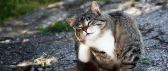
Irritation/Red Eyes in Dogs
Red eyes in dogs are a common symptom of many eye diseases. The cause could be something as simple as an allergy. However, it can also be caused by infection, corneal ulcers, chronic dry eye, tumors, glaucoma and a variety of other problems.
Contact your veterinarian if your dog has red eyes. Your veterinarian will be able to determine the cause by other symptoms, including eye discharge, squinting, and corneal clouding.
Squinting in dogs
Squinting is a sign of eye pain in dogs. This is common in many eye conditions, including allergies, infections, corneal ulcers, chronic dry eye syndrome and glaucoma. It may also be associated with painful eyelid lesions such as entropion, tumors and skin infections.
If you see your dog squinting, take him to the vet.
Glass eyes in dogs
Glassy eyes are a symptom of eye inflammation, which can be caused by pink eye, chronic dry eye, or allergies. Dehydration also causes glassy eyes.
Solving the problem depends on identifying the cause and treating it. So, if your dog has glassy eyes, is lethargic, isn't eating, or isn't acting normally, take your dog to the vet as soon as possible.
Cloudy eyes in dogs
There are many causes of cloudy eyes in dogs:
- Corneal ulcers
- Chronic dry eye
- Inflammation of the cornea due to infection
- Diseases of the eyelids that cause irritation of the cornea, such as entropion or eyelid tumors.
- Glaucoma and eye tumors
A veterinarian should examine cloudy eyes as soon as possible to determine the cause and prescribe treatment.
Resolving the problem quickly will make your dog more comfortable and reduce the risk of permanent eye damage.
Dull eyes in dogs
All dogs have a small amount of clear or white discharge in the corners of their eyes; This is fine. The color and amount of eye discharge are important clues to what eye disease your dog may be suffering from.
Some dog breeds tend to produce more tears than others, causing the inner corners of the eyes to turn a rust color. Watery, clear discharge is typical with allergies or blocked nasolacrimal ducts. Yellow or green discharge is characteristic of a bacterial infection. Very thick and dry discharge that sticks to the eye is usually associated with chronic dry eye syndrome.
If you notice discharge from your dog's eyes, contact your veterinarian.
Height/bump on a dog's eyelid
Eyelid tumors are very common in middle-aged and older dogs.
Benign tumors on the edge of the eyelid, called meibomian cysts, are usually not a problem unless they are large enough to rub against the surface of the eye. If the cyst becomes inflamed or large enough to damage the dog's eye, surgery will be required to remove it.
There are malignant tumors that can occur on the eyelids, such as mast cell tumors, squamous cell carcinoma, and melanoma, and these will require surgical removal.
Contact your veterinarian to check for any swelling on your dog's eyes.
Third eyelid visible in dogs
A dog's third eyelid is an important structure: it holds the largest tear gland in the eye and protects the eye.
When the lacrimal gland falls out (displaces itself), it is visible in the third eyelid as a large, smooth, red mass on the upper edge of the third eyelid. Tumors may occur in the third eyelid, including lymphoma, melanoma, hemangiosarcoma, and squamous cell carcinoma. Tumors make the third eyelid larger and more noticeable.
Nervous conditions such as Horner's syndrome and tetanus can cause the eye to retract into the socket, causing the third eyelid to move into the visual field.
Eye pain also causes the dog to retract the eye deeper into the socket, which can also cause a third eyelid to appear. A wrinkled eye and dehydration are two other causes of a visible third eyelid.
Contact your veterinarian if your dog's third eyelid is visible.
Lens luxation
Lens luxation (luxation) - the corresponding part of the eye is displaced from the hyaloid fossa. Lens luxation in a dog can be partial or complete.
Cause. Lens luxation in a dog can be due to genetic predisposition, glaucoma, cataracts, and as a result of severe injuries and infectious diseases suffered by the dog. Lens luxation occurs in dogs as a result of rupture of the ligaments of the lens and the ciliary muscle. Terriers are more susceptible to this disease.
Symptoms. During a clinical examination of a dog with a similar pathology, a veterinarian notes a deformation of the pupil, its displacement away from the center or it is swollen, and the shape of the eyeball itself may change. There is a disruption in the movement of fluid in the ocular body.
Treatment. Treatment of lens luxation is carried out in a veterinary clinic through surgical correction. After removal of the lens, an intraocular lens implant is placed. In especially valuable dogs, it is possible to implant the entire eyeball.
Glaucoma
Glaucoma or “green star” is a group of diseases associated with increased pressure inside the visual apparatus. They are characterized by redness of the eye, significant enlargement of the pupil, and a greenish cataract. During the course of the disease, photophobia, apathy, enlargement of the eyeball, and slower pupil reaction are observed. The problem is hereditary, and certain breeds are genetically predisposed to it.
Treatment does not give a 100% result, but it can slow down development. First of all, they reduce the pressure inside the eye. Then they use the cyclocryotherapy method: cold to the ciliary body. Medicines prescribed for pets are aimed at improving the outflow of intraocular fluid.
| List of drugs for treatment | ||
| Name of medicine | Price | Release form and application |
| Azopt | 947 rubles per bottle (5 ml) | Eye drops that suppress fluid production by almost half |
| Alphagan | 718 rubles per bottle (5 ml) | Drops that reduce pressure inside the eye and increase the outflow of fluid; apply 2-3 times a day, 1-2 drops |
Any disease can lead to injury or atrophy of the eye, but individual diseases of the mucous membranes also occur. It is important for the owner not to get confused and start treatment on time: contact a veterinarian, start using medications.
4.7 / 5 ( 3 voices)
Eyeball luxation
When the eyeball is dislocated, dog owners note that the eyeball comes out of the orbit behind the eyelid completely or partially.
This pathology is most often found in Pekingese, Japanese hips and similar breeds of dogs.
Cause. Dislocation of the eyeball in a dog most often occurs due to mechanical damage to the bones of the head and temples, high muscle tension in dogs with a shallow depth of the bone orbit.
Clinical picture. During a clinical examination, the veterinary specialist of the clinic notes a strong extension of the eyeball beyond its natural boundaries, the conjunctiva is swollen, often dries out, and externally takes the form of a hanging cushion.
Treatment. The treatment of this pathology is surgical.
Pathologies of the fundus and lens
This group includes diseases that are predominantly irreversible. The development of pathology often causes the pet’s eyes to become cloudy and also affects the quality of vision.
Retinal atrophy
At this stage of development of veterinary medicine, it is believed that the disease is of hereditary origin. At an early stage, the dog's twilight vision deteriorates and night blindness develops. Further development of the disease gradually affects the acuity of daytime vision, gradually leading to complete blindness. The color of the affected eye is pale.
Retinal detachment
This process may be a natural consequence of atrophy. In addition, trauma and neoplasms, accompanied by an increase in blood pressure, can lead to retinal tissue detachment. In this case, vision loss occurs more rapidly, and hemorrhages are observed on the surface of the eyeball.
Cataract
A disease that affects the lens and causes it to become cloudy. “Gray star” can be either hereditary or acquired. It often joins the course of hormonal diseases such as diabetes, as well as retinal diseases. No effective treatment has been found.
Recommended Posts
Why do dogs have red whites of their eyes and should they be concerned?
Typical diseases and vaccinations of the Shih Tzu breed
Symptoms and treatment of diseases in pugs
Table and calculations for determining the age of a cat by human standards
What and how to properly wipe a kitten’s eyes
Lens diseases
Cataract is a disease of the lens accompanied by partial or complete opacity of the lens and its capsule.
Cataracts in dogs can be primary. In which a veterinarian, during a clinical examination, notes isolated damage to the eye area or systemic diseases in the animal.
In Boston Terriers, West Highland White Terriers, and Miniature Schnauzers, cataracts may have a hereditary form.
Primary juvenile cataract is considered the most common form of cataract in all breeds of dogs and mixed breeds. It is usually registered in dogs under 6 years of age.
Secondary or sequential cataracts in dogs are non-inherited cataracts.
Congenital cataracts usually occur in dogs together with other congenital eye changes.
Acquired - occurs in dogs with retinal diseases, eye abnormalities in collies, injuries, diabetes.
Retinal detachment and atrophy
A hereditary disease caused by eye abnormalities, injuries, high blood pressure - detachment. The disease is characterized by a rapid course. Sudden hemorrhage, severe blindness, and impaired pupil reflex occur. Retinal detachment is practically incurable. Requires surgery and antibacterial drugs.
Atrophy is a hereditary genetic problem. It manifests itself as a slow decrease in the quality of vision: first in the dark, then in daylight, complete blindness and clouding of the pupil. The disease is incurable.