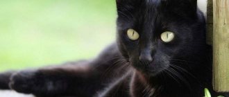
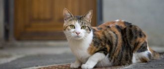
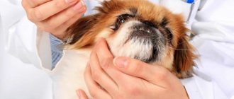
Often, a dog owner notices that his pet is “crying.” Flowing tears leave wet marks on the animal's face and color the corners of the eyes brownish or reddish.
Why do my dog's eyes water? Most often this is due to the breed characteristics of the animal or foreign particles getting into the eyes, but sometimes the problem can be more serious and require the help of a veterinarian.
Causes
Normally, dogs may leak a little tears, but if the tearing becomes too profuse and bothers your pet, then this is a signal that the process has gone beyond what is natural.
Tear fluid has antibacterial properties and helps protect and moisturize the cornea of the eye. With the help of tears, all foreign objects and pathogenic microorganisms are removed from the surface of the eyeball.
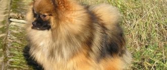
Some dog breeds are characterized by excessive lacrimation, this is due to the size of the animal or the anatomical features of the development of the skull and eyes. Thus, tears often flow from the eyes of small dogs, for example, toy terriers, due to their size.
A short muzzle with a flattened nose is also a cause of eye discharge in Pugs, Shar-Peis and other breeds. From this we can conclude that if you have a puppy of this breed, he will have a predisposition to lacrimation.
But, if your dog’s eyes are too watery, the reasons for this may be:
- pollution with sand, dust, debris;
- allergies to food, household products;
- owners smoking;
- irritation of the eyeball by hair or eyelashes;
- entropion of the eyelid;
- problems with the lacrimal canal - an inflammatory process in it, blockage or a congenital anomaly of the canal;
- eye injury;
- infectious diseases.
In the case of pathological lacrimation, the volume of tear fluid increases, it becomes opaque, and this process causes discomfort to the dog.
Lens luxation
Luxation , or luxation of the lens , is a serious disease in which the lens is displaced due to rupture of the suspensory ligaments (ciliary band) into the anterior (most often) or posterior chamber or into the vitreous body, or it is pinched in the pupil.
Symptoms:
- Pain
- Photophobia
- Tearing
- Squinting (blepharospasm)
- There may be a violation of the shape of the pupil
Treatment:
1. Immediate surgical removal of the lens, because secondary complications often develop - glaucoma and uveitis. The earlier the luxated lens is removed, the better the prognosis.
===========================================================================================================================================================================================
How to cope on your own
- If the cause of discharge from your dog’s eyes is a foreign object or an aerosol that has entered the eyes, then you can help your pet yourself. To do this, you need to thoroughly rinse your eyes with warm boiled water, boric acid solution or chamomile infusion. A cotton swab, generously soaked in the solution, is drawn from the outer edge of the eye to the inner, in the direction of the nose. Each eye is wiped with a separate swab. Such manipulations must be carried out several times a day for up to 5 days in a row.
- Tearing associated with eyelashes or fur located near the eye does not require treatment. In this case, you need to carefully trim excess hair or too long eyelashes. This will have to be done constantly so that the dog does not experience discomfort.
- In a too dry room, your pet's eyes may also water, so you need to humidify the air or instill special drops such as “Artificial Tear” in your dog, which help nourish and lubricate the cornea.
- Special care will also be required for animals of those breeds for which excessive production of tears is the norm. In this case, you need to take care of the dog’s eyes, wiping them daily with boiled water so that the accumulated tear fluid does not become a breeding ground for pathogenic microflora. This type of care should continue throughout your pet's life.
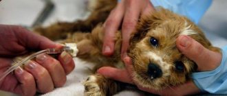
Professional treatment
In a veterinary clinic, before starting therapy, the doctor will definitely find out the cause of the disease. This may require taking blood tests, calculating the leukemia formula, and additional studies are possible in each individual case.
Owners often ask whether it is possible to drip Albucid into the dog’s eyes. Human medications are not recommended to be given without consulting a veterinarian. An ophthalmologist, indeed, can in some cases prescribe this drug, but only after examining the patient. After all, if used incorrectly, albucid can cause irritation of the mucous membrane and cause a chemical burn. Your veterinarian may also prescribe special veterinary drops. They contain recommended dosages of medications and are safe to use.
If the visit to the veterinarian was late, purulent inflammation can provoke the appearance of a cataract. Cloudiness of the cornea is accompanied by the appearance of a white spot on the dog’s eye. In this case, an appointment with an ophthalmologist is required. And to establish a final diagnosis, an ultrasound of the eye, examination of the fundus, and measurement of intraocular pressure may be required. If a complication is confirmed —
the necessary therapy is prescribed. You cannot delay this, because untimely treatment can result in loss of vision for your pet.
Symptoms
My dog’s eyes have been watering for a long time, what should I do? If you have already tried to treat your pet at home, and it did not help, then it is better not to delay time and go to the veterinarian.
You should be wary if:
- Eye washing has been carried out for several days, but there is no improvement in the condition;
- eyes turned red;
- the discharge changed color and consistency, the eyes began to turn sour;
- the dog's eyes became cloudy;
- the dog squints its eyes, tries to scratch them, blinks frequently;
- the dog began to see worse;
- The general condition of the dog worsened.
If such symptoms are observed, it is necessary to urgently contact a veterinarian so that he can carry out a qualified examination and prescribe appropriate treatment.
Cataract
Cataract is clouding of the lens, leading to a decrease in visual acuity, up to its complete loss. The clouding may be partial or complete. The release of protein from the lens (phacolysis) leads to chronic phacolytic uveitis (endophthalmitis), a severe intraocular inflammation. The disease can be congenital or hereditary and can occur at any age.
Symptoms:
— Decreased or loss of vision in the dog, blurred eyes.
Treatment:
Currently, no conservative (therapeutic) methods for treating cataracts have been developed. To restore visual function and treat secondary complications, surgical treatment is effective, during which the affected lens is removed and, if indicated, an artificial lens is implanted.
============================================================================================================================================================================================
Diseases accompanied by lacrimation in dogs
If you are sure that your pet does not have a foreign object in his eye and there has been no trauma, then why are his eyes watering?
The cause of lacrimation can be infectious diseases, inflammatory eye diseases and problems with the tear ducts:
- Lack of exit from the nasolacrimal canaliculus - congenital or acquired as a result of a previous disease.
- Stenosis of the nasolacrimal duct.
- Inflammation in the lacrimal sac, which occurs against the background of narrowing of the nasolacrimal duct.
- Turning of the eyelid inward, causing irritation of the eyeball by eyelashes.
- Blockage of the tear ducts.
- Neoplasms and inflammatory processes in the system that removes tears.
- Allergic reaction to external irritants or food.
- Infectious diseases.
- Conjunctivitis of various etiologies.
- Inflammation of the cornea of the eye - keratitis.
- Inflammation of the vascular network of the eye - uevitis.
- Glaucoma.
All these diseases are quite dangerous for a dog, and untimely and incorrect treatment can not only worsen your pet’s vision, but even completely deprive it of it.
Adenoma of the third century
Adenoma of the third eyelid , or correctly said – prolapse (prolapse) of the lacrimal gland of the third eyelid.
Usually observed in young dogs aged from 6 months to 2 years, most often in brachiocephalic breeds (Shih Tzu, cocker spaniels, pugs, bulldogs, etc.), beagles, etc. This occurs due to genetically determined weakness of the ligaments that fix the gland to the edge of the eye socket. Symptoms:
— An oval red formation appears in the inner corner of the eye, protruding from under the edge of the third eyelid. It is most often observed in one eye, but can also be bilateral.
- There may be moderate lacrimation and squinting of the eyes (blepharospasm).
Treatment:
1. The gland must be returned to its place surgically.
============================================================================================================================================================================================
How to treat
What to do if the cause of your dog's lacrimation is related to a disease? If this is a general disease of an infectious nature, then the tears will stop flowing as soon as the underlying disease is cured.
For inflammatory eye diseases - conjunctivitis, uevitis, keratitis, complex treatment is prescribed, including anti-inflammatory, antibacterial drugs, which are applied to the eye in the form of drops or ointments. If necessary, veterinarians recommend additional medications in tablets or injections.
The most popular drugs used to treat conjunctivitis, keratitis, and blepharitis in dogs are veterinary drops - “Tsiprovet”, “Bars”. They contain antibiotics that are active against gram-positive and gram-negative microorganisms and also have an anti-inflammatory effect.
Glaucoma in dogs has practically no treatment, only drugs are prescribed that lower intraocular pressure and alleviate the animal’s condition.
For lacrimation associated with pathologies of the lacrimal system, surgical intervention is most often necessary. During the operation, the patency of narrowed, overgrown or blocked areas is restored, after which tear drainage begins to occur normally.
Watery eyes in a dog suffering from allergies due to poor diet or exposure to external factors will return to normal after the animal’s contact with the allergen stops. Additionally, the doctor may prescribe antihistamines.
To preventively moisturize the surface of a dog’s eye in a dry room, you can use “Diamond Eye” or “Optimmune” drops.
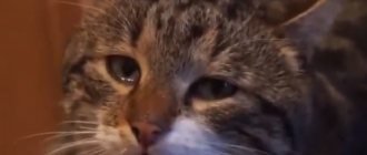
Etiology - why the eyes turn red
“Red eyes” is the most common clinical symptom noted by pet owners. Redness of the eyes as a clinical symptom cannot indicate the severity of the disease, its type or prognosis. The first diagnostic step is to localize the redness. It can be focal and generalized, external and internal and extend to the conjunctiva, third eyelid, periscleral region and cornea. When a patient is brought in with the main complaint of a red eye, the cause of the redness is first identified, then it is determined whether the animal has superficial or internal eye damage. Sources of eye redness include:
- conjunctival vessels;
- scleral vessels;
- cornea;
- front camera;
- neovascularization of the iris.
“Red eye” may not affect the health of the animal in any way, but with some diseases associated with it, vision may weaken or disappear, the eye may be destroyed, and sometimes the disease can cause the death of your pet.
Conjunctivitis in a dog
Redness indicates a superficial or deep pathological process. Internal eye pathologies most often require urgent treatment and can lead to blindness in the absence of appropriate treatment. The superficial ones include the conjunctiva and superficial vessels of the cornea. Among the internal causes of redness are the vessels of the sclera, the anterior chamber of the eye and neovascularization of the iris.
| Sources of eye redness | Causes | Symptoms and diagnostic tests |
| Vessels of the conjunctiva. Their noticeable injection indicates a superficial lesion. | Keratoconjunctivitis sicca (KSK), dystrichiasis, primary corneal disease or irritation | The vessels are thin and branched, and move with the conjunctiva. To differentiate between conjunctival vasodilation and scleral vasodilation, a drop of phenylephrine or epinephrine is applied to the affected eye. The vessels of the conjunctiva constrict within 15 seconds. |
| Scleral vessels. Vasodilation indicates internal damage to the eye and can lead to blindness developing within 24-48 hours if the primary disease is not treated. | Anterior uveitis, glaucoma, scleritis, deep corneal lesions | The vessels of the sclera are thick, straight and not branched. They do not move with the conjunctiva and do not narrow when a vasoconstrictor is administered. |
| Vascularization of the cornea. May indicate superficial or internal disease depending on the location of the vessels. | Superficial vessels indicate nonspecific irritation (eg, ulcers, foreign body, SBS). Deep vessels are a nonspecific indicator of the response to a deep pathological process (scleritis, deep scleral disease). | Superficial vessels cross the limbus and are often long and branching. Deep vessels: short and straight with brush-like edges. It is not visible how they cross the limbus, since they are located deep. All lie to one certain depth. |
| Anterior chamber - hyphema | The same causes and pathological picture as with bleeding in other areas of the body | These include: trauma, tumor, hypertension (more common in older dogs with kidney disease), uveitis, coagulation disorders, retinal detachment, secondary neovascularization (the retina secretes substances that cause neovascularization. These new vessels may have increased permeability). |
| Neovascularization of the iris | Chronic uveitis, lymphosarcoma | In dogs with a lightly pigmented iris, a large arterial ring is normally visible. |
Differential diagnosis of red eye based on additional characteristics:
- state of visual function. With conjunctivitis, vision is not affected. On the contrary, patients with glaucoma often experience serious vision problems, and even blindness. Anterior uveitis does not cause blindness, but the patient may have corneal edema or aqueous opacification, aqueous hyperemia, hyphema, or hypopyon, which can also affect vision;
- pain reaction. The acute period of glaucoma is very painful: with the occurrence of blepharospasm and general depression. The chronic stage of the disease is also extremely painful, but changes in behavior are less pronounced. Similarly, severe anterior uveitis may be moderately painful, but chronic uveitis shows no signs of pain. Conjunctivitis is painless, or irritation is moderate;
- presence of outflows. Anterior uveitis and glaucoma are often accompanied by increased lacrimation. Conjunctivitis is characterized by serous, mucoid or purulent discharge from the eye.
- condition of the conjunctiva . With conjunctivitis, the conjunctiva is thickened, diffusely hyperemic and edematous. In glaucoma and anterior uveitis, it has a normal consistency.
- injection of blood vessels. It is important to establish what causes the redness: congestion in the vessels of the conjunctiva, episcleral region or ciliary body. Mobilization of the conjunctiva by flushing will cause its vessels to move, but will not affect the condition of the deep vessels. For example, topical application of phenylepinephrine causes immediate discoloration of the vessels of the conjunctiva, but practically does not affect the vessels of the episclera and ciliary body. The redness of conjunctivitis is most obvious on the surface of the eyelids and arch of the eye. The vessels are usually narrow, and diffuse stagnation is observed. The episcleral vessels, in which congestion occurs in glaucoma, are much wider and are visible on the bulbar surface towards the border. The vessels of the ciliary body, stagnant in uveitis, are indistinguishable because they are located deep. But they make the eye red. Please note that both glaucoma and anterior uveitis can contribute to the appearance of congestive processes in the vessels of the conjunctiva, as well as in the episcleral and ciliary vessels.
- hypertrophy of lymphatic follicles with conjunctivitis; Glaucoma can cause band keratopathy (stretching and thinning the sclera), as well as retinal and optic disc atrophy. In glaucoma, excavation of the optic disc is pathognomic;
- Anterior uveitis is accompanied by loss of transparency of the aqueous humor. This may manifest as aqueous hyperemia, hyphema, hypopyon, or cellular debris on the anterior lens capsule and posterior cornea. As a result, anterior or posterior synechia may form;
- Anterior uveitis can spread to the back of the eye, leading to posterior uveitis and inflammation of the ciliary body. In severe cases, optic neuritis/atrophy and endophthalmitis occur.
Red whites of eyes
The most common cause of redness in the whites of the eyes in dogs is allergies. Most often, an allergy that manifests itself as discharge from the eyes and conjunctivitis occurs to environmental components (pollen, dust, etc.), and an allergy that occurs on the skin is often due to flea bites. Secondly, allergies occur to food components.
Doberman Pins and Great Danes are especially susceptible to redness of the whites of the eyes.
Less commonly, proteins acquire a red color due to chronic eye pathologies. Chronic disease of the cornea can lead to vascularization with possible granulation of corneal tissue. Hyphema can fill the entire chamber, then the entire cornea will be red and completely cover the iris and pupillary opening. Bleeding in the vitreous can also cause redness of the eyes (this should be distinguished from the normal red fundus reflection in dogs). A retinal detachment may appear as blood vessels against the caudal side of the lens.
Lump in the corner of a dog's eye
The disease is accompanied by redness and thickening of the edges of the eyelids, scales, crusts and ulcers appear at the base of the eyelashes. Eyelashes fall out, the edges of the eyelids become very thick, which leads to constant tearing and cicatricial inversion.
Follicular conjunctivitis is a chronic non-infectious inflammation of the mucous membrane of the eyes, which affects its lymphatic follicles. Mechanical irritants include all kinds of injuries, foreign bodies entering the conjunctival sac, inversion and eversion of the eyelids. All this directly irritates the conjunctiva or disrupts its protection from harmful external influences - dust, smoke, prolonged exposure to direct ultraviolet and x-rays
The occurrence of this pathology is due to the loose structure of connective tissue and subcutaneous tissue. Most often, hypertrophy and prolapse of the third eyelid gland occurs in dogs of such breeds as:
- mastino;
- bullmastiffs;
- Great Danes;
- English bulldogs;
- chow-chow;
- Shar Pei.
Inflammation of the third eyelid in dogs, noticeable to humans, can occur in connection with various ailments. So, with prolapse (loss) of the third eyelid, its protective shell is no longer held in place by the thin ligament that holds the gland in the orbit of the eye. Due to prolapse of the third eyelid, it can be further injured, which is fraught with complications. A similar phenomenon often occurs in dogs aged 3 to 9 months during a period of rapid growth.
During this period, dogs also experience such a pathology as a third eyelid crease: the thin cartilaginous leg that holds the eyelid breaks off, and the eyelid hangs over the eye as in the previous case. It is also worth noting that third eyelid adenoma in dogs can be caused by other diseases: disorders of the central nervous system, traumatic injuries to the cornea, atrophy of the eyeball, and others. After localizing the redness, a full examination of the eye should be performed to diagnose concomitant diseases, give a prognosis and prescribe treatment. To do this, the pupillary reflex, blink reflex, intraocular pressure are determined, the intraocular fluid is examined, the cornea is stained with fluorescein, the Schirmer tear test, and the eyelids, cornea, iris, lens, vitreous body and fundus are carefully examined.
Additional tests to determine the cause of eye redness:
- Gauss test, excluding corneal rupture;
- Detection of damage to the cornea (staining it with fluorescein);
- Measurement of pressure inside the eye;
- Analysis of eye secretions using a microscope;
- Assessment of the functioning of the lacrimal glands;
- Ultrasound of the eye to clarify the overall picture;
- X-ray of the skull;
- Computed tomography of the skull;
- Magnetic resonance imaging of the brain.
Canine distemper virus often causes conjunctivitis with epiphora, mucous or purulent discharge. Along with systemic symptoms, keratoconjunctivitis sicca, corneal ulceration, chorioretinitis and optic neuritis may also occur. Cytological examination of scrapings from the third eyelid gland usually reveals cytoplasmic inclusions. Direct or indirect immunofluorescence antibody testing can also be performed.
Prevention
Prevention involves daily examination and care of your pet's eyes.
What symptoms should you be wary of:
- The eye waters for a long time;
- The discharge is mucous, dense, thick (not watery);
- Tears are not transparent;
- Smudges are normally brownish, dark, if they are of a different color - pathology;
- The eye is red, swollen, itchy.
To examine the mucous membrane of the eye, place your thumb on the edge of the eyelid and pull it down or up. Normally it is pale pink, moderately moist.