Everyone knows earthworms; they constitute a large group of different species belonging to the family Oligochaetes.
The common earthworm belongs to the most famous family Lumbricidae, consisting of approximately 200 species, and about 100 of them are found in our country. The body length of the common earthworm reaches 30 centimeters.
Earthworms (Lumbricina).
External structure
Body
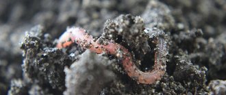
The earthworm, or earthworm (Fig. 51) has an elongated body, 10-16 cm long. In cross section, the body is round, but, unlike roundworms, it is divided into 110-180 segments by annular constrictions.
Each segment has 8 small elastic bristles. They are almost invisible, but if we run our fingers from the back end of the worm's body to the front, we will immediately feel them. With these bristles, the worm rests against uneven soil or the walls of the passage when moving. Regeneration in earthworms is well expressed.
Body wall
If we take a worm in our hands, we will find that its body wall is wet and covered with mucus. This mucus makes it easier for the worm to move through the soil. In addition, only through the moist body wall does the oxygen necessary for breathing penetrate into the worm’s body.
The body wall of an earthworm, like that of all annelids, consists of a thin cuticle, which is secreted by a single-layer epithelium.
Under it there is a thin layer of circular muscles, and under the circular muscles there are more powerful longitudinal muscles. By contracting, the circular muscles lengthen the body of the worm, and the longitudinal muscles shorten it. Thanks to the alternating work of these muscles, the worm moves.
Subtleties of proper care
To extend the lifespan of earthworms, they need to be properly cared for, providing comfortable conditions for development and reproductive functions. The key rules of care are to ensure a comfortable temperature, shade and stable access to food.
Professional gardeners recommend enriching compost manure with sand or crushed eggshells. Dry food is added to the soil approximately once every two weeks, but it is important to take precautions here so as not to overfeed the worms.
If you want to start breeding creatures at home, you need to remember that worms process almost everything that catches their eye. The main thing is that the products are small in size, because invertebrates do not have teeth.
Before replenishing the worm house with new food, you need to make sure that the previous organic matter is completely destroyed. If this is not the case, the animals will suffer from oversaturation and die. Leaving organic compounds in the compost will negatively impact the acidity levels, leading to deadly conditions. In addition, excess food supply contributes to the development of pests such as mites.
Caring for worms is relatively easy, but some requirements must be met. Only under such conditions will breeding a home farm bring the expected success.
Habitat
Earthworm and its movement in the soil. The anterior end of the earthworm's body from below.
During the day, earthworms stay in the soil, making tunnels in it. If the soil is soft, the worm penetrates it with the front end of its body. At the same time, he first compresses the front end of the body so that it becomes thin, and pushes it forward between the lumps of soil. Then the front end thickens, pushing the soil apart, and the worm pulls up the rear part of the body.
In dense soil, the worm can eat its way through the soil through its intestines. Lumps of soil can be seen on the surface of the soil - they are left here by worms. After heavy rain has flooded their passages, the worms are forced to crawl out to the surface of the soil (hence the name - earthworm). In summer, worms stay in the surface layers of the soil, and in winter they dig burrows up to 2 m deep.
Arrangement of the worm house
If you want to start breeding earthworms at home, you need to equip a spacious worm barn in advance. The structure should become a comfortable home for the spineless, where they can live, eat and perform their biological duties.
A wooden box, a trough, an old bathtub and other similar structures can be used as a container. Experienced gardeners recommend placing high-quality open compost in the container. However, the ground must be protected with netting to prevent mass consumption of eukaryotes by birds and other natural enemies.
Proper worm care involves preparing suitable fertile soil. About 40 centimeters of compost should be placed at the bottom of the box, after which it should be filled with warm water. Next, you should create a straw bedding and wait a few days until the substances are completely absorbed. After completing these steps, you can put the worm into operation.
Finding the first group of worms to move is not difficult. It is enough to go to your own garden or vegetable garden and dig up a small layer of soil. Individuals living in the upper layers tolerate movement into the worm nest better than all others. Invertebrates are also sold in specialized gardening stores.
Settlement is carried out in several stages. The first thing a gardener needs to do is dig a small hole and then place a bucket of worms there. After this, the animals are covered with straw or burlap. In a week you will be able to notice the first results of the worms taking root.
To prevent the death of the colony, it is necessary to regularly monitor them. If the creatures demonstrate an active lifestyle, it means that they have managed to settle down normally , and everything is fine with them.
To improve adaptation to the new environment, the worms need to be fed only after 3-4 weeks. However, the heated liquid should be placed in the worm chamber at least twice a week.
Digestive system
Internal structure of an earthworm.
Digestive, nervous, circulatory system of an earthworm. The mouth is located at the anterior end of the earthworm's body; the anus is on the back.
The earthworm feeds on rotting plant debris, which it swallows along with the soil. It can also drag fallen leaves from the surface. Food is swallowed as a result of contraction of the muscles of the pharynx. The food then enters the intestines. Undigested remains, along with soil, are expelled through the anus at the rear end of the body.
The intestines are surrounded by a network of blood capillaries, which ensures the absorption of nutrients into the blood.
Methods for controlling nematodes
To combat root nematodes, insecticides and biological preparations are used. Traditional methods of struggle are ineffective. Herbal decoctions and infusions do not help. You can plant plants containing phytoncides on the site - calendula, marigolds, mustard salad, garlic. They can repel pests.
It is best to carry out preventive measures in the fight against nematodes, namely:
- compliance with crop rotation;
- cultivation of varieties resistant to the pest;
- use of healthy planting material;
- regular liming of the soil;
- mandatory destruction of plant residues at the end of the season;
- sowing green manure.
Such measures will prevent the appearance of the pest in the soil and help preserve the harvest.
The carrot fly is a small insect with an oblong black body and is unremarkable. Its larvae damage carrots, parsnips, root parsley and celery.
When affected, the leaves of the plant acquire a purple-red hue, gradually turn yellow and dry out. Root vegetables lose their taste and shelf life.
Effective agrotechnical measures include early sowing of carrots and other root crops sensitive to the pest and immediate removal of affected plants. During the growing season, vegetable crops can be treated with insecticides or biological preparations.
“Corado” from carrot and onion flies
Preventive measures include:
- compliance with crop rotation;
- alternating crops when sowing (it is better to plant carrots in areas where garlic, onions, tomatoes, and radishes previously grew);
- when sowing, alternate rows of carrots with rows of onions or garlic;
- thin out seedlings in a timely manner;
- do not allow the soil to become waterlogged.
It is thanks to preventive measures that you can save plants and get a full harvest.
The onion fly is one of the types of flower flies, the larvae of which damage onions, garlic and bulbous flower crops. The insect is small, yellowish-gray. The larvae are white, up to 10 mm long. They penetrate into the plant from the bottom or through the base of the leaves. They eat the bulb from the inside. The leaves of damaged plants turn yellow and wither, and the bulb itself rots. Some larvae are able to migrate to neighboring bulbs.
Onion fly larvae eat the onion
Early planting or sowing of onions can prevent onion fly damage. When pests are identified, diseased plants must be immediately destroyed, and the remaining ones must be treated with an insecticide.
As part of prevention, you should:
- follow the rules of crop rotation, replanting onions on the site no earlier than after 4 years;
- pickle planting material with manganese and copper sulfate;
- treat plants with repellents (tobacco, hot pepper, ash);
- use soil insecticides (for example, Bazudin).
It is also allowed to treat plants with insecticides or biological agents during the growing season, but provided that the onions are not grown for feathers.
You should not lose sight of the animals that live in the ground and, during their activity, cause no less damage to vegetable and garden crops than insects.
Circulatory system
All secondary cavities have a circulatory system, starting with annelids. Its occurrence is associated with a mobile lifestyle (compared to flat and protocavitary worms). The muscles of annelids work more actively and therefore require more nutrients and oxygen, which the blood brings them.
The earthworm (Fig. 52) has two main blood vessels: the dorsal, through which blood moves from the rear end of the body to the front, and the abdominal, through which blood flows in the opposite direction. Both vessels in each segment are connected by ring vessels.
Several thick annular vessels are muscular; due to their contraction, blood moves. Muscular vessels (“hearts”) located in segments 7–11 push blood into the abdominal vessel. In the “hearts” and spinal vessels, valves prevent blood from flowing back.
From the main vessels, thinner ones depart, which then branch into the smallest capillaries. These capillaries receive oxygen through the surface of the body and nutrients from the intestines. From the capillaries that branch in the muscles, carbon dioxide and decay products are released.
Blood moves all the time through the vessels and does not mix with the cavity fluid. Such a circulatory system is called closed. Blood contains hemoglobin, which is able to carry more oxygen; she is reddish.
A closed circulatory system allows you to significantly increase your metabolic rate. In annelids it is twice as high as in flatworms, which do not have a blood pumping system.
Whales in the sand
Having borrowed the body from worms and the mouthparts from lampreys, the great worms of the sandy oceans of Arrakis took the method of feeding from completely different creatures - whales. They feed on sandy plankton, filtering it out like baleen whales on Earth.
Many filter-feeding organisms known to us reach significant sizes (baleen whales, whale sharks, manta rays), although they live in water. In earth's deserts you can find animals that literally “swim” in the sand, but all these creatures are relatively small in size.
Scincus scincus , covered with smooth scales, “swims” well in the sand.
and sand boas
Eryx miliaris
, pharmacist skinks are even called “sand fish”.
Excellent sand swimmers are found among golden moles and marsupial moles. Golden moles (family Chrysochloridae
) - relatives of tenrecs and otter shrews, they got their name for the golden color of their coat with a metallic tint.
the golden mole Eremitalpa granti
alternates between “swimming” in the sand and walking along the surface of the sand, during which it immerses its head in it and listens to vibrations.
In addition to insects, golden moles also hunt skinks (they share different habitats with skinks) and are nicknamed “sand sharks.” Marsupial moles of the genus Notoryctes
have an amazing convergent similarity in lifestyle and appearance with golden moles.
Filter-feeding organisms, strictly speaking, do not need teeth, although there are exceptions. The whale shark Rhincodon typus
about three thousand teeth, although small and unsuitable for biting anything.
We don’t know how the great worm’s filtration occurred, but it is known that Arraki sand is primarily a product of the digestion of sandworms. Probably, the feeding method of the great worms was closer to the feeding method of earthworms, which simply swallow soil fragments (and any other organic matter) and throw back the processed mass in the form of coprolites. But among aquatic vertebrates there is an example of substrate filtration: for example, gray whales Eschrichtius robustus
They are the only whales that can filter silt.
On the other hand, in the Encyclopedia of Dune we find information that diverges from the original text of Dune that the great worms are autotrophs. Let us recall that autotrophs are organisms that synthesize organic substances from inorganic ones. That is, according to the Encyclopedia, in addition to feeding on sandy plankton, great worms also use certain inorganic compounds; the sand itself may also be used in metabolism. At the same time, when the great worms move through the thickness of the sand, their covers rub against it, and the contact generates electricity and heat, which is also involved in the metabolism of the great worms. On Earth, we do not know examples of such simultaneously huge and mobile autotrophs.
Reproduction and development
Earthworms are hermaphrodites. During the process of copulation of two individuals, mutual fertilization occurs, that is, the exchange of male gametes, after which the partners separate.
The ovaries and testes are located in different segments at the anterior end of the body. The location of the reproductive organ system is shown in Figure 51. After copulation, a belt is formed around each worm - a dense tube that secretes the cocoon shell.
The cocoon receives nutrients that the embryos will subsequently feed on. As a result of the expansion of the rings located behind the cocoon, it is pushed forward towards the head end.
At this time, 10-12 eggs are laid through the opening of the oviduct into the cocoon. Further, when the cocoon moves, sperm from the sperm receptacles received from another individual during copulation enters it, and fertilization occurs.
After this, the cocoon slides off the worm and its holes quickly close. This prevents the eggs it contains from drying out. The development of earthworms is direct, that is, they do not have larvae; a young worm hatches from the egg.
Earthworm coming out
To lean out of the hole and throw out excrement, the worm extends its tail forward, and if the worm needs to collect leaves, it sticks its head out of the ground. That is, in burrows, earthworms can turn over.
Earthworms do not always throw out soil near the surface; if they find a cavity, for example, in plowed soil or near the roots of trees, then they throw out excrement into this cavity. There are small clumps of earthworm excrement between many rocks and under fallen tree trunks. Sometimes worms fill their old holes with excrement.
№11
They are quite voracious creatures. They can eat anywhere from ½ of their body weight to their entire body weight each day. For example, if we compared their gluttony with a person, then a person with a body weight of 70 kg could eat from 35 to 70 kg of food per day.
Each earthworm can process about 4.5 kg of organic material per year.
Parascariasis in horses
Parascariasis is a helminthiasis in which parascaridae are localized in the small intestine, and sometimes in the stomach and bile ducts of the animal's liver. Parascariasis is observed everywhere. This disease mainly affects horses.
Parascarid eggs in the external environment reach the invasive stage in 1-2 weeks. From the moment a horse ingests parasite eggs until their sexually mature stage, approximately 1.5-2 months pass.
Equine parascariasis is more common in places with a humid climate and marshy soil. Mostly young individuals in stable and mixed housing are susceptible to the disease. Adult horses get sick less often.
Infection of horses with parascaridae is more often recorded in the autumn-winter period, after which a decrease in the disease is noted.
Horses are infected through the nutritional route by ingesting invasive parasite eggs in feed or water. Foals can become infected with parascariasis while nursing mares.
The symptoms of this disease, caused by nematodes, depend on the intensity of the damage, the age of the animals, the resistance of their body and possible allergic reactions.
The symptoms are most pronounced in foals, while in adults parascariasis is mainly subclinical. When parascarid larvae migrate through the blood in the first days after infection, enteritis and diarrhea are observed, then bronchopneumonia develops, accompanied by a cough, a short-term increase in body temperature and mucous discharge from the nose. In the presence of parascaridae in the intestines, digestive upset occurs, animals quickly lose weight and get tired quickly. The mucous membranes become pale, the composition of the blood changes, the number of red blood cells and hemoglobin decreases. Leukocytosis, eosinophilia, and accelerated erythrocyte sedimentation reaction are often observed. Sometimes foals experience convulsions or paresis.
To diagnose this disease, it is necessary to examine horse feces using the Fulleborn method. In foals infected with parascariasis, parascarid eggs cannot always be detected, so diagnostic deworming is recommended.
Piperazine, carbon disulfide and carbon tetrachloride are used to treat parascariasis.
Piperazine is indicated in the form of piperazine sulfate, piperazine hexahydrate in a dosage of 8-25 g per head (depending on the age of the animal) twice for 2 consecutive days with food or water.
Carbon tetrachloride should be administered in a dosage that depends on the age of the animal. For foals from 3 to 7 months, this drug is indicated in a dosage of 5-10 ml, from 7 to 12 months - 10-15 ml, from one year to 2 years - 15-20 ml, for adult horses - 25-40 ml. In case of significant helminth infestation, it is recommended to give a saline laxative after carbon tetrachloride. Before administering the drug, animals are given a 16-18-hour fasting diet. During the deworming period, concentrates should be completely excluded from the diet. Manure must be removed daily and disinfected using the biothermal method. Approximately 10 days after deworming, the stable should be treated with a hot 5% carbolic acid solution.
For prevention, scheduled deworming of horses should be carried out in the fall and spring. It is prohibited to feed roughage to horses from the floor. Animals can be given water only with clean drinking water.
It is possible to defeat parasites!
Antiparasitic Complex® - Reliable and safe removal of parasites in 21 days!
- The composition includes only natural ingredients;
- Does not cause side effects;
- Absolutely safe;
- Protects the liver, heart, lungs, stomach, skin from parasites;
- Removes waste products of parasites from the body.
- Effectively destroys most types of helminths in 21 days.
There is now a preferential program for free packaging. Read expert opinion.
Read further:
What worms look like in children: roundworms, pinworms, flukes and tapeworms
The largest worm: tapeworms, roundworms and earthworms
Roundworms in humans and domestic animals: what they look like and how to treat them
Worms in herring (round, tapeworms): are they dangerous for humans, is it possible to eat and what to do about it
What cat worms look like: what worms do cats get and how to recognize them
Class tapeworms: characteristics of representatives and their lifestyle
Spirocercosis in dogs
Spirocerciasis is a disease of dogs caused by roundworms that parasitize in inflammatory tubercles of the esophagus, stomach, aorta and lungs.
Beetles act as intermediate hosts and eat eggs released by sick animals. It is in the body of the beetles that the parasite larvae reach the invasive stage.
Dogs become infected by eating infested beetles.
There are reservoir hosts (birds, reptiles, etc.) that feed on beetles. The larvae remain in their body for a long time and serve as a source of infection for dogs. In the dog's body, the larvae travel with the blood to the localization sites, around which swellings appear with a hole at the top, from where the eggs of mature helminths come out.
Helminths reach sexual maturity in about 5 months.
The rapid spread of the disease is due to the presence of a wide range of reservoir hosts.
The presence of swelling in the aorta, esophagus, stomach and other vital organs significantly impairs the properties of these organs. Symptoms of the disease reflect the pathology of those organs in which the parasites are localized, therefore, disturbances in the rhythm of blood flow, swallowing, coughing, etc. are observed. Diagnosis of this disease requires a set of studies, including laboratory ones. Fecal examination is carried out using the Fulleborn method. To detect swelling, fluoroscopy of organs can be performed.
No specific therapy has been developed, but levamisole and ivermectin can be used for treatment. Animals are injected intramuscularly with a 10% aqueous solution of levamisole at a dosage of 5 mg/kg for 2 days. A 1% solution of ivermectin is indicated as a subcutaneous injection at a dosage of 0.02 ml/kg. The course of treatment is 3-5 days.
To prevent spirocerciasis in dogs, animal feces should be regularly disposed of by burning or burying.
Setaria in farm animals
Setariasis is a helminthiasis that is caused by nematodes that parasitize in the abdominal cavity (mature individuals), in the brain and spinal cord, in the eyes (juvenile forms) and in the blood (microsetaria).
Many types of farm animals are susceptible to this disease, including cattle, sheep, goats, and horses.
Setaria develop with the participation of intermediate hosts (mosquitoes and flies). The development of the parasite in the definitive host occurs within 6 months. Juvenile forms of helminths parasitize in the brain and spinal cord of sheep, and its mature forms are localized in the abdominal cavity of cattle.
Infection occurs by inoculation of infective larvae by blood-sucking insects on pasture. The greatest number of infections occurs during the flight of insects.
The main symptoms of this nematode, caused by setaria nematodes, depend on the degree of damage. In horses and cattle, clinical manifestations have not been studied. Paresis and paralysis of the limbs are observed in sheep. When the brain is damaged in sheep, movement coordination disorder occurs, which often leads to the death of the animals.
To diagnose setaria in horses and cattle, it is necessary to conduct a blood test to detect microsetaria.
No special therapy has been developed. Control and prevention measures include preventing contact between definitive and intermediate hosts. It is important to combat insects that carry setaria.
Parafilariasis in horses
Parafilariasis is a disease of horses in which helminths parasitize the subcutaneous tissue of animals. The causative agents are white thread nematodes.
The course of this disease is characterized by seasonality. Basically, the first cases of infection occur in April, and the peak incidence occurs in July-August. The higher the air temperature, the more pronounced the disease manifests itself. The largest number of horses become infected as a result of prolonged stay on pastures. Most often adults become ill.
Helminths, moving in the subcutaneous tissue, actively drill into the skin of the animal. In places of lesions, tubercles form on the skin of infected horses. Over a few days they gradually increase to the size of a pea. They appear mainly on the withers and shoulder blades, in the back and ribs, on the neck, and sometimes on the lower back and croup. Then, in these places, in sunny weather, holes and wounds form, through which blood leaks from broken capillaries and small vessels. At night the bleeding stops. After some time, the wounds heal and the tubercles disappear.
Bleeding is not observed daily throughout the summer season, but intermittently at different times. There can be more than 150 bleeding wounds on a horse's body. With such intensity of helminth damage, the horse loses a large amount of blood. When a collar or saddle comes into contact with wounds on the horse's skin, phlegmon appears.
In places where parasites live, abscesses that arise as a result of inflammation of the subcutaneous tissue can form without external influence.
To establish a diagnosis, it is necessary to take into account the seasonal characteristics of the invasion, as well as the symptoms of this type of nematode. It is recommended to conduct a blood test from emerging wounds in order to detect eggs and larvae of nematodes. To do this, a drop of blood on a glass slide should be diluted with 10 drops of distilled water and, without covering it with a coverslip, placed under a microscope. Blood can be drawn into a centrifuge tube and diluted with water in a ratio of 1:10, and the resulting sediment can be placed under a microscope. There are no microfilariae in the circulating blood, so there is no need to distinguish them from microsetaria.
To treat this disease, it is recommended to inject a weak aqueous solution of potassium permanganate subcutaneously into the affected areas in a ratio of 1:1000.
Ivomec should be administered subcutaneously at a dosage of 1 ml/50 kg or ivermectin at a dosage of 0.2 mg/kg.
To prevent parafilariasis, it is recommended to use various insecticides.