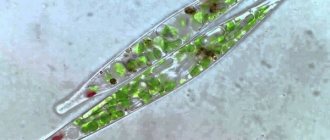
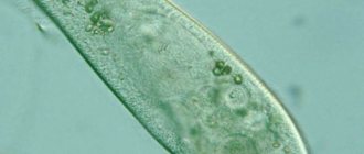
Euglena protozoa are single-celled flagellated organisms, the most famous representative of which is Euglena green.
Many textbooks indicate that green euglena has one flagellum, but in fact there are two of them (like most euglenaceae): one long locomotor flagellum, clearly visible in a light microscope, and the second very short and therefore invisible.
They feed by phagocytosis or diffusion. Many euglenoids have chloroplasts and synthesize their own nutrients through photosynthesis, without losing the ability to also absorb food from the environment (mixotrophic nutrition).
Euglena movement
Most species of euglena prefer to swim in the water column and move with the help of flagella. However, there are species that live on the substrate on which they have adapted to crawl. They have very short flagella that do not take part in movement, and movement is carried out due to cell peristalsis - wave-like bending of the cell body, or metabolism.
Metabolic movement is characteristic only of euglenoids, which is why it is also called euglenoid. It is a kind of crawling movement. Its mechanism and biophysics are not fully understood. Apparently, it is provided by protein ribbons underlying the membrane, which run in a spiral along the entire body and can slide past each other.
Euglena intermedia moving by metabolism. A short flagellum is visible that does not take part in movement. Microscope magnification 1000x
In harsh, bright light, the metabolic movement of euglena can acquire a very large amplitude.
Intense peristalsis indicates that the euglena does not like too bright light (it can destroy chlorophyll), and by contracting, it tries to reduce its impact:
Metabolic movement accelerated 10 times
Ciliate slipper
In fresh water bodies, the slipper ciliate is often found, which got its name due to the peculiarities of the cell shape. Her body is shaped like a shoe. This protozoan moves with the help of cilia.
Among freshwater protozoa, one shoe has the most complex structure. Inside the ciliate there are two nuclei at once: large and small. The large nucleus regulates all life processes, the small one plays an important role in the reproduction of the shoe.
Ciliates feed on bacteria, algae and some other protozoa. With the help of vibrations of the cilia, she pushes food into the mouth, and then into the pharynx. Digestive vacuoles form at the bottom of the pharynx, where food is digested and nutrients are absorbed. Undigested residues are removed through a special organ - powder.
The reproduction of the slipper ciliate is influenced by its more complex structure compared to other protozoa, since it has two nuclei. One is large, called a macronucleus, the second is small, called a micronucleus.
The slipper ciliates have not only asexual reproduction, but also sexual reproduction. However, it proceeds far from the same as in multicellular animals. With it, paradoxically, the number of individuals does not increase.
Two different cells of the ciliate slipper are involved in the sexual process. They approach each other from the side of the cell mouths and stick together. A so-called cytoplasmic bridge is formed between them - a channel through which the contents of one cell can flow into another. This process of reproduction is called conjugation .
Thanks to this process, the genetic material inside the ciliate cells is updated, and it becomes possible for new traits to arise that can contribute to their better survival in the environment.
Asexual reproduction of the slipper ciliate proceeds in approximately the same way as in amoeba and green euglena. The cell divides in two. However, unlike the same euglena, ciliates are divided not in the longitudinal direction, but in the transverse direction. That is, one daughter cell gets the front part of the cell, and the second gets the back part.
Phototaxis. How does the light-sensitive eye of euglena work?
For a long time it was believed that the stigma, a red pigment spot at the base of the flagellum, was responsible for photosensitivity. But then it turned out that stigma does not have photoreceptors and does not have photosensitivity
However, according to tradition, it is still called the photosensitive eye, and this name continues to wander from textbook to textbook.
In fact, it is only part of the photoreceptor apparatus. Many flagellates that do not have stigma can respond successfully to light.
The light receptor is a section of the membrane at the base of the flagellum, located directly under the pigment spot (if present).
Why then do we need stigma?
Euglena moves by rotating along its longitudinal axis. The stigma and its position during rotation are clearly visible. Microscope magnification 1000x
As can be seen in the video, when swimming, the euglena rotates around the longitudinal axis, respectively, one day a situation will arise when the rays of light either directly hit the receptor or the stigma obscuring it. Changing the illumination of the photoreceptor affects the functioning of the flagellum, which allows it to move towards the light. That. The stigma acts as a screen and helps to effectively determine the direction of light. However, its functions are not limited to just a shutter; it also works as a reflector and as a diffraction grating, i.e. converts the light signal and provides a stronger change in its intensity.
Example of phototaxis
In the video below, euglena, following the call of phototaxis, gather in the most illuminated place - in the center of the preparation.
But even a good thing can be too much. Too much light can damage the chloroplasts, and the euglena then evacuate the danger zone together, leaving the middle empty.
Phototaxis of euglena. Microscope magnification 400x and 200x
Nutrition
The nutrition of Euglena Green is not only half autotrophic, but half heterotrophic. A suspension of starch-like substance accumulates in the cytoplasm of the cell. This is a nutritional reserve for a rainy day. The mixed type of nutrition is called mixotrophic by scientists. If Euglena gets into bodies of water hidden from light, for example, caves, it gradually loses chlorophyll.
Then the unicellular alga begins to look more like a simple animal, feeding exclusively on organic matter. This once again confirms the possibility of kinship between plants and animals. When there is lighting, the heroine of the article does not resort to “hunting” and is sedentary. Why wave a flagellum if food itself falls on you in the form of light? Euglena begins to move actively only in twilight conditions.
The algae cannot survive the night without food because it is microscopic. There is simply nowhere to make sufficient energy reserves. The accumulated amount is immediately spent on vital processes. If Euglena is starving, experiencing both a lack of light and a lack of organic matter in the water, it begins to consume a starch-like substance. It is called paramil. Animals also use fat stored under the skin.
The protozoan Euglena green to a backup method of nutrition , as a rule, in a cyst. This is the hard shell that the algae forms when compressed. The capsule is like a bubble. Actually, the concept of “cyst” is translated from Greek.
Before cyst formation, the alga discards its flagellum. When unfavorable conditions are replaced by standard ones, the cyst germinates. One Euglena, or several, may emerge from the capsule. Each grows a new flagellum. During the day, Euglenas rush to well-lit areas of the reservoir, staying near the surface. At night, single-celled organisms are distributed over the entire area of a pond or river backwater.
Red Euglena (Euglena sanguinea)
Euglena sanguinea at 400x and 1000x microscope magnification
. Not all euglena are green. Since ancient times, the bloody euglena Euglena sanguinea has terrified people with its ability to instantly turn bodies of water blood red. Then, of course, they did not know about the existence of any euglena and perceived the mass flowering of these protozoa as a bad omen or as a harbinger of God's punishment.
This euglena in its normal state is almost no different from its famous relative, green euglena. But when the light is too bright, it quickly turns red, using the carotenoid pigment astaxanthin as protection from too intense solar radiation. This very pigment is used on fish farms, where it is added to feed to color the meat of salmon fish and crustaceans.
In the video you can see granules of this pigment, and green chloroplasts peeking out from under them:
Red euglena surrounded by green euglena of another species. Microscope magnification 1000x
Euglena algae: Euglenophyta
Unicellular motile forms with one or two flagella, sometimes without them; the cells are naked, the role of the shell is played by the outer layer of protoplasm, sometimes the cell is in a “house”.
Most algae are green in color, sometimes light green due to the presence of xanthophyll. They reproduce by longitudinal cell division. Representatives of euglena algae are euglena and facus.
Euglena genus
- Euglena (Fig. 250). Euglena lives in small fresh water bodies, on damp soil, in brackish waters, and in rice fields.
Sometimes, multiplying in huge quantities, it causes red, brick-red or green blooming of water. It feeds phototrophically or mixotrophically. Reproduces by cell division. The genus is rich in species (more than 60). Euglenas are found in reservoirs of the European part of the USSR, Western Siberia, Crimea, Central Asia, Estonia and Latvia.
They participate in the self-purification of waters and serve as food for protozoa and insect larvae.
They live inside the intestines of various animals. Some euglena serve as an indicator of water pollution by organic substances.
Rice. 250 Euglena and red algae: 1 - euglena; 2 - focus; 3 - batrachospermum
Rod faculty
- Phacus (Fig. 250). The algae is widespread in shallow reservoirs among thickets of aquatic plants, in the plankton of lakes and ponds, often together with euglena.
It feeds phototrophically and mixotrophically. Reproduces by cell division. Quite widely distributed in water bodies of the USSR. Sometimes it causes algae in water. More than 40 types of facus are known.
The most likely time for their abundant appearance in the aquarium is summer.
Trachelomonas
Trachelomonas at 1000x microscope magnification
Trachelomonas is a relative of Euglena green, a flagellate living in a house called a lorica.
The lorica has a hole surrounded by a neck, from which a long flagellum emerges. You can see the chloroplasts and the red eye - stigma.
It reproduces by binary fission, after which one of the individuals leaves the house through the hole and immediately synthesizes a new lorica for itself. A newly built house is absolutely transparent and colorless. Over time, it acquires a brown tint due to the deposition of iron and manganese salts.
The simplest, but not simple
“The nervous system of a bug no larger than a pin demonstrates spontaneity; even an amoeba has its whims, its follies!”
Stanislav Lem
The body of unicellular organisms consists mainly of the nucleus and cytoplasm. It also contains mitochondria, ribosomes, the Golgi apparatus and other organelles characteristic of plant and animal cells. In the cytoplasm of protozoa there are digestive and contractile vacuoles. They are responsible for digesting food and removing waste from the body. Almost all protozoa are capable of active movement. They move using pseudopods, flagella and cilia.
GIANTS OF THE WORLD OF PROTOZOTS
The largest protozoa are considered to be marine rhizomes of foraminifera. Their length was 15-20 centimeters. These animals became extinct about 70 million years ago.
Most protozoa feed on bacteria and decaying organic matter. Among them there are species with autotrophic, heterotrophic or mixed types of nutrition. Autotrophs have chromatophores - organelles containing photosynthetic pigments. Heterotrophic protozoa absorb ready-made organic substances from the environment.
Single-celled creatures live in fresh water bodies, seas and soil. Most of them know how to wrap themselves in a dense cocoon - a cyst - when unfavorable conditions occur. As soon as conditions become favorable, the animal leaves its sealed capsule and begins to feed and reproduce as before.
Let's look at how the simplest organisms are structured, using the example of three prominent representatives: amoeba, green euglena and slipper ciliates.
Euglena Lepocinclis fusca
Euglena Lepocinclis fusca looks thorny because... has an ornament on the pellicle in the form of tiny pyramids.
In the video you can see orderly rows of triangular structures.
Also clearly visible is a red light-sensitive eye and two large oval structures - grains of the reserve nutrient paramyl. You can see a short flagellum that does not take part in movement. It is carried out by bending the cell.
Euglena Lepocinclis fusca at 1000x microscope magnification
Amoeba
“The path from amoeba to man seems to some to be an obvious progression, but it is not known whether amoeba agrees with this opinion.”
Bertrand Russell, British philosopher
Amoeba is a freshwater representative of the rhizome class. Unlike many protozoa, it does not have a constant body shape. Its only cell is constantly transforming. The amoeba moves with the help of pseudopods.
The pseudopods also serve to capture food - bacteria, unicellular algae and some of the simplest relatives of the amoeba. Having grabbed the victim with its pseudopods, the amoeba seems to swallow it. The prey ends up in the cytoplasm, where a digestive vacuole forms around it. Under the influence of digestive juice coming from the cytoplasm, food is digested. Nutrients penetrate into the cytoplasm, and undigested residues are thrown out.
AMOEBA - PROTEUS
Amoeba was first described and drawn by the German naturalist Johann Rösel von Rosenhof. He named her Proteus - in honor of the ancient Greek god who changes his appearance at will. Since then, “amoeba” has been a symbol of something simple, artless and narrow-minded.
Amoeba reproduces by division. The nucleus divides in two, both halves diverge, and a constriction forms between them. Then two daughter cells arise from one mother cell. After the division process is completed, they continue to live independently, independently of each other.
Colacium
Euglena genus colacium on crustacean.
Microscope magnification 200x and 400x On crustaceans, you can often find ciliates in the role of epibionts (i.e., organisms that live on the surface of other organisms), but in this case they are euglena protists Colacium. They are the only genus of euglena that lead an attached lifestyle, settling on small planktonic animals.
In their development cycle there is a motile stage during which they have a flagellum and look like a typical euglena, but when they find a host, the flagellum is shed and a mucous stalk is formed for attachment to the body of the animal.
Department of euglena algae
The department includes microscopic unicellular organisms equipped with one or two flagella and actively moving. The body shape of euglena algae is elongated, oval, ellipsoidal or fusiform. There is no cellulose casing; its role is played by the outer compacted layer of cytoplasm - the pellicle. Those species in which the pellicle is soft and elastic have the ability to change the shape of the body.
Few algae have an outer hard shell, usually impregnated with iron salts, which does not adhere tightly to the protoplast. The number and shape of chromatophores are different.
Order Volvocida
Plant flagellates with 2–4 flagella and a cup-shaped chromatophore. They live in marine and fresh water bodies. There are both solitary (Chlamydomonas) and colonial forms (Gonium, Eudorina, Volvox, Pandorina) (Fig. 2).
rice. 2. Colonial flagellates. A - gonium, B - eudorina, C - volvox.
Volvox globator is a freshwater planktonic colony of flagellates; the cells of the colony are called zooids (Fig. 2B). There are vegetative and generative zooids, both of which are haploid. The colony has a spherical shape. The bulk of the colony is a gelatinous substance formed as a result of mucilage of the cell walls. Vegetative zooids are located along the periphery of the colony and are connected to each other by cytoplasmic bridges. Generative zooids determine reproduction. In spring, generative zooids plunge into the mother colony and divide by mitosis, forming daughter colonies. Daughter colonies, after the destruction of the mother colony, move on to independent existence. In autumn, macrogametes and microgametes are formed from generative zooids. The diploid zygote, formed as a result of copulation of gametes, overwinters, divides by meiosis in the spring, and new colonies are formed from haploid zooids.
►