- Signs
Euglena green is the simplest single-celled organism, unique in that there is still no unanimous agreement among biologists as to which kingdom it belongs to, animals or plants. The fact is that green euglena combines equally the characteristics of both plants and animals. Since euglena contains chlorophyll, during the day it is powered by sunlight through the process
photosynthesis, exactly as all other plants do, but at night, in the dark, it transforms: with an abundance of organic food, it can feed heterotrophically, that is, as all animals do. Euglena green is also capable of moving, again, like all other animals. It is believed that green euglena is a transitional form from plants to animals; its existence confirms the theory of the unity of all living things. And according to this theory, man descended not only from monkeys, but also from plants, so trees and flowers are our distant relatives, but let’s return to euglena, what is its structure, habitat, what does it eat, how does it reproduce, read on.
Description and characteristics. What does green euglena look like?
The body of green euglena consists of twenty
chloroplasts, which contain chlorophyll, which is involved in photosynthesis. Chloroplasts are green plates and in general they are only present in cells with a nucleus in the center. And thanks to them, euglena is green and is called “green”, due to chloroplasts and chlorophyll it is really bright green.
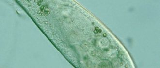
This is what green euglena looks like when you look at it under
microscope.
If during the day euglena receives energy from sunlight through the process of photosynthesis, then at night it feeds on organic matter from the water. The water itself must be fresh. Therefore, euglena is found in fresh water bodies: ponds, lakes, rivers, swamps.
In appearance, euglena is similar to algae, and would be such a unicellular algae, if not for several nuances. Firstly, heterophoric night feeding of euglena is characteristic of animals, but not plants. In addition, there are other signs that euglena belongs to animals:
ciliates are slippers, whose movements are always smooth due to the large number of small cilia.
Due to all these points, there is still no consensus in the scientific community about where Euglena green belongs: to plants or animals. Most scientists still classify it as a flora, seeing it as a unicellular algae, 15% of biologists consider it an animal, the rest see it as an intermediate species.
Features of green euglena
Euglena green is an algae. It got its name due to its green color. A special feature of the cell is its versatility. It can be either autotrophic or heterotrophic: it is able to feed on everything that is available to it. It is characterized by a high chlorophyll content, which allows it to convert sunlight into a source of nutrition. This property allows it to be classified as a plant.
If the lighting is very weak, it switches to the second type of nutrition - heterotrophic. Due to the fact that it has been in the dark for a long time, chlorophyll is lost and the microorganism becomes white. The main nutritional organ in the heterotrophic type is the vacuole.
And the more neglected the reservoir, the more nutrients it contains.
Signs
The body of our heroine is spindle-shaped with a hard shell. The average body length of euglena is 0.5 mm. The front part of the body is blunt and has a red eye. This eye is photosensitive and allows its owner to find “feeding” places during the day, in other words, “it leads the euglena to the light.” In any body of water, these microorganisms always gather in the brightest places. By the way, a large number of euglena in a given body of water makes the surface of the water reddish, even brown. Such an unusual effect from the accumulation of euglena was observed and described in his works by the great naturalist of antiquity, Aristotle, in the 4th century BC. e.
At the anterior end of the body of a unicellular organism there is a flagellum. Moreover, in newborn organisms the flagellum may be absent, since the cell is divided into two and the flagellum remains on only one of the parts. On the second euglena it will grow back over time.
The rear end of the green euglena's body, on the contrary, is pointed; this shape improves streamlining, and therefore speed.
Interestingly, green euglena is characterized by metabolism, that is, the ability to change body shape. Despite the fact that, as a rule, euglena are fusiform, in different circumstances they can take other forms, be:
- like a cross
- rolled,
- spherical,
- lumpy.
But regardless of the body shape of the green euglena, its flagellum will be invisible if the cell is alive. And he is invisible for the reason that the frequency of his movements is so fast that the human eye is simply not able to catch it.
Functions of green euglena
To reach the brightest area. To determine the most convenient place, she has a peephole at the very throat. It is very sensitive and is able to respond to any changes in light. The process of constantly moving in search of light is called phototaxis. To carry out the process of osmotic regulation, euglena has contractile vacuoles. They allow you to get rid of any excess substances. The vacuole received this name because it has the property of contracting, thereby all processes occur faster.
Euglena has only one set of chromosomes. In addition to chloroplasts, the cytoplasm contains paramyl (this is a reserve protein).
The microorganism also has a core and separate areas with inclusions of nutrients. They will help you survive the period when food is difficult. In protozoa, breathing occurs completely with the whole body.
Euglena adapts to any, even the most severe, conditions. If conditions become worse and food runs out, the cell becomes rounded and is enveloped in a special membrane to protect it from external influences. The flagellum of the cell disappears. Euglena can remain in this position for a long time.
Structure
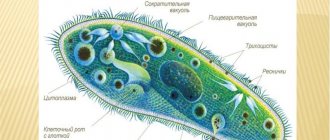
Summarizing all of the above, we can conclude that green euglena is an animal or plant consisting of:
- A flagellum, the very presence of which places our heroine in the class of flagellates. The diameter of the flagellum is on average 0.25 micrometers; it can only be seen through a powerful microscope. The process is covered with a plasma membrane consisting of microtubules that move relative to each other. Their movement causes the general movement of the flagellum.
- Eye, also sometimes called stigma. The eye consists of optic fibers and lens-like formations. Thanks to the latter, it captures the light that the lens reflects onto the flagellum. Having received an impulse from it, the flagellum, in turn, begins to move towards the light. The red color of the euglena eye is due to colored droplets of lipid - fat. The peephole itself is surrounded by a membrane.
- Chromatophore are special pigmented cells and plant components responsible for its color; in euglena they are bright green.
- Peplicula is a Latin word that means “skin.” Euglena peplicules, consisting of flat membrane vesicles, form the shell of this simplest single-celled organism.
- A contractile vacuole, which is located just below the base of the flagellum. This contractile vacuole is a kind of analogue of muscle tissue. In the structure of the euglena, it is responsible for pushing out excess water from the cell, due to which the euglena maintains its constant volume.
This is how the structure of green euglena looks in the picture.
A few more words about the contractile vacuole, with its help the respiration of green euglena is also carried out.
Euglena green flagella
The flagellum is an important element of the cell, thanks to which it is able to move. The flagellum is also important in the feeding process. Its structure is quite simple: a small protrusion from the cell. Thanks to rapid movement, the microorganism is able to move and capture nutrients.
When viewed under a microscope, the flagellum performs rotational screw movements. It’s like he’s screwed into his environment. The movement occurs at a very high speed and creates a whirlpool, as a result of which small nutrient particles are attracted to the cell. A vacuole is formed at the base of the flagellum, in which nutrients are digested.
Habitat
Euglena lives only in fresh water bodies, especially preferring those where the water is dirtier. In reservoirs with clean water, euglena is either small in number or completely absent. In this respect, euglena is similar to its other single-celled “colleagues”: amoebae and ciliates, which also love dirty water.
Since euglena are quite resistant to cold, in addition to fresh water they can live in harsh conditions of ice and snow.
It is worth noting that green euglena can be dangerous, as living in putrefactive water, it sometimes serves as a carrier of trypanosomes and leishmania. The latter is the causative agent of some skin diseases. Trypanosomes can cause African sleeping sickness, which affects the nervous and lymphatic systems, leading to fever.
If euglena gets into aquarium water, then such water will bloom, so it is not without reason that aquarists consider euglena a dangerous parasite and try to get rid of it. You can get rid of green euglena using special chemicals (remembering to move the fish to another place during this time). And, of course, do not forget about regular water replacement and filtration, then the water in the aquarium will be fresh and clean and euglena will not grow in it.
Behavior of green euglena
When observing a euglena under a microscope, its bold and bold behavior is visible. She is constantly on the move and strives to capture more and more territory.
When left for a long time without light, the body of the microorganism becomes colorless. This is the result of a lack of photosynthesis. In such situations, a single-celled microorganism switches completely to a heterotrophic mode of nutrition.
Euglena travels great distances. The cell is assisted in this by a flagellum, which accelerates its movement with a screw-like rotational movement.
Nutrition
As we wrote above, the nutrition of this creature is half heterotrophic and half autotrophic, that is, both due to solar energy and due to organic matter. This unusual, mixed type of nutrition, characteristic exclusively of the life activity of green euglena, is called mixotrophic by biologists.
During the day, euglena is under the Sun, it is not in a hurry and inactive, and really, why should it move and wave its flagellum if “food” in the form of sun rays falls on you itself. But if a euglena finds itself in some dark reservoir hidden from the Sun, and also at night, then it transforms from a plant into an animal, its flagellum begins to actively move, moving its owner around the reservoir in search of organic “food”.
Therefore, if during the day euglena are located only in the light parts of the reservoir, and usually close to the surface of the water, then at night they spread throughout the entire reservoir.
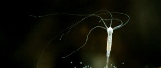
Red Euglena (Euglena sanguinea)
Euglena sanguinea at 400x and 1000x microscope magnification
. Not all euglena are green. Since ancient times, the bloody euglena Euglena sanguinea has terrified people with its ability to instantly turn bodies of water blood red. Then, of course, they did not know about the existence of any euglena and perceived the mass flowering of these protozoa as a bad omen or as a harbinger of God's punishment.
This euglena in its normal state is almost no different from its famous relative, green euglena. But when the light is too bright, it quickly turns red, using the carotenoid pigment astaxanthin as protection from too intense solar radiation. This very pigment is used on fish farms, where it is added to feed to color the meat of salmon fish and crustaceans.
In the video you can see granules of this pigment, and green chloroplasts peeking out from under them:
Red euglena surrounded by green euglena of another species. Microscope magnification 1000x
Organoids
Organelles or organelles are the permanent or specialized structures of every cell, both animal and plant. As for the organelles of green euglena, they have already been listed above, in the section on the structure of euglena. Each of these organelles or organelles is a vital element of a single-celled organism, without which it would not be able to eat, move, reproduce and generally exist.
Euglena green cytoplasm
The fluid that fills the euglena is called cytoplasm. The main substance in its composition is hyaloplasm. It consists of proteins, polysaccharides and nucleic acids. Compounds similar to starch accumulate in it. They practically float in the water. This environment is the cytoplasm. The ratio of components in its composition is constantly changing and does not have clear standards. All elements appear colorless. Euglena's green color comes from chlorophyll.
Reproduction
Would you like, dear reader, to live forever? This is a philosophical question, and you may be surprised, but in biology there is an example of “infinite life”, and yes, our today’s heroine, Euglena, is this example. The lifespan of green euglena is essentially endless! And all because of the method of its reproduction, which is carried out exclusively through cell division. So the euglena that you can see today in some green pond or swamp was created through division from a certain euglena that lived in the era of dinosaurs, or even earlier.
But the time that euglena remains indivisible is, on the contrary, extremely short, amounting to only a few days. Then the euglena begins to divide, then divide again, and so on ad infinitum.
As for the division of euglena itself, it occurs in several stages, it all begins with the division of the cell nucleus. Two new nucleoli diverge on opposite sides of the cell, after which the cell itself begins to divide in the longitudinal direction. Cross division is not possible.
This is how the division of euglena looks schematically.
The divided membrane closes on each half of the cell. Thus, from one euglena you get two. In a favorable environment, these creatures can reproduce in arithmetic progression.
Euglena green kernel
The nucleus is the basis of all single-celled organisms. It is located in the tail part of the body, it is held by chromatin threads. Thanks to the division of the nucleus, euglena reproduces.
After division, two new nuclei are repelled in different directions of the cell, then the microorganism itself is separated into two longitudinal parts. Transverse division never occurs. The rupture occurs along the fission line of new nuclei. After division, the membrane comes together at the break points, resulting in two separate cells. Euglena green is able to reproduce in all states of liquid (water, snow and ice).
Recommended reading and useful links
- Green euglena is a kind of flagellate. Volvox // Biology: Animals: Textbook for grades 7-8 of secondary school / B. E. Bykhovsky, E. V. Kozlova, A. S. Monchadsky and others; Edited by M. A. Kozlov. — 23rd ed. - M.: Education, 1993. - P. 14-16. — ISBN 5090043884.
- Biology: handy. for 8th grade, dark lighting. navch. concluding / S. V. Mezhzherin, Ya. O. Mezhzherina. - K.: Osvita, 2008. - 256 p. ISBN 978-966-04-0617-9.
- Mikheeva T. M. Euglena // Belarusian Encyclopedia: U 18 t. T. 18. Book. 1.: Dadatak: Shchytniki - YA. - Mn. : BelEn, 2004. - T. 18. - P. 186. - 10,000 approx. — ISBN 985-11-0295-4 (Vol. 18. Book 1.).
Colacium
Euglena genus colacium on crustacean.
Microscope magnification 200x and 400x On crustaceans, you can often find ciliates in the role of epibionts (i.e., organisms that live on the surface of other organisms), but in this case they are euglena protists Colacium. They are the only genus of euglena that lead an attached lifestyle, settling on small planktonic animals.
In their development cycle there is a motile stage during which they have a flagellum and look like a typical euglena, but when they find a host, the flagellum is shed and a mucous stalk is formed for attachment to the body of the animal.
Video
Author: Pavel Chaika, editor-in-chief of Poznavaika magazine
When writing the article, I tried to make it as interesting, useful and high-quality as possible. I would be grateful for any feedback and constructive criticism in the form of comments on the article. You can also write your wish/question/suggestion to my email [email protected] or Facebook, with respect, the author.
Author page
Trachelomonas
Trachelomonas at 1000x microscope magnification
Trachelomonas is a relative of Euglena green, a flagellate living in a house called a lorica.
The lorica has a hole surrounded by a neck, from which a long flagellum emerges. You can see the chloroplasts and the red eye - stigma.
It reproduces by binary fission, after which one of the individuals leaves the house through the hole and immediately synthesizes a new lorica for itself. A newly built house is absolutely transparent and colorless. Over time, it acquires a brown tint due to the deposition of iron and manganese salts.
Trichomonas
A genus of flagellated protozoa, three species of which parasitize humans: T. vaginalis (vaginal Trichomonas, parasitizes the genitourinary tract, causing intractable diseases), T. tenax (oral Trichomonas) and T. hominis (intestinal Trichomonas, causes chronic diarrhea).
In its development cycle, Trichomonas has three stages: flagellated (adult stage), amoeba-like (intermediate and most aggressive), cyst-like (can exist in a special shell that protects it from harmful external influences) and many transitional forms.
The flagellated trophozoite phase has 4-6 flagella, one of which is a steering flagellum and forms an undulating membrane. Infection with Trichomonas can occur through the mouth, rectum, genitals, or by inhaling air that contains these flagellates.
Type Euglenozoa
The phylum unites about 1000 free-living, parasitic and epibiont (living on the surface of the body of other organisms) species, distributed throughout the world. Among them there are both photoautotrophic (green) and heterotrophic (colorless) protists. Most autotrophic euglenids live in fresh water bodies. They predominate in shallow, standing waters with a large amount of organic matter. Developing in large quantities, they can cause green and red “blooming” of water.
Flagellates - Euglena
Heterotrophic euglenids are also found in the ocean shelf zone. Most of them have 2 heterokont (unequal in length) flagella. There are species with one and several flagella. The surface of the euglenid cell is covered with a cuticle, often with a highly developed layer of glycocalyx; some species have houses encrusted with iron salts.
The movement is flagellar or euglenoid (metabolic) . Metabolic movement is characteristic of species living on the surface of the substrate, and not inside it; it occurs by elongation and compression of the cell, its expansion, folding and bending. Such protists may lack a flagellum.
Species capable of photosynthesis have a photoreceptor that includes a paraflagellar body and a stigma. The paraflagellar body is an expanded section of a long flagellum covered with a membrane. The interaction of the photoreceptor and the flagellar apparatus leads to a change in the direction of movement of euglenids. Stigma is red or orange-red, which is due to the concentration of carotene in it.
They have chromatophores In the cells of different euglenids there can be from one to several hundred. Many have pyrenoids - differentiated areas inside the chromatophores of algae and photosynthetic protozoa, in which enzymes that synthesize reserve sugar (paramylon) are concentrated.
The mitochondrial cristae are lamellar; in anaerobic euglenoids, mitochondria are absent. Everyone has only one nucleus in the cell; it is divided according to the type of closed pleuro- or orthomitosis. The main method of reproduction is cell division in two; in some species, palintomy - a process of successive divisions without stages of growth and increasing the volume of the resulting cells.
The life cycle usually contains forms for surviving unfavorable conditions: palmelliformes (mucous accumulations of divided cells) and cysts . The shape of the actively reproducing stage ( trophozoite ) of this protozoan can be leaf-shaped, spherical, ellipsoidal, ovoid, needle-shaped, fusiform.
Feeding methods of euglenids:
- holophytic;
- saprozoic;
- holozoic;
- mixotrophic.
In the absence of light, many green species become colorless. They feed heterotrophically and require an organic carbon source. Various changes occur in their cell: pyrenoids, chloroplasts, and often stigmas disappear. When moving towards the light, their chloroplasts and other organelles are restored and photosynthesis begins again.
There is only one contractile vacuole in the cell. Its contents are poured into the reservoir of the flagellar apparatus, and from there it is thrown out. The reserve storage substance of all euglenids is the polysaccharide paramylon . In green individuals, the center of its formation is the pyrenoid; in heterotrophs, it is produced in other parts of the cytoplasm. Paramylon is deposited in the cytoplasm in the form of grains of different sizes - paramylia .
In heterotrophic colorless species, in addition to paramylon, glycogen and deposited in the form of small grains. The reserve substance of euglenids is also neutral fat , deposited in the form of drops. The blood-red euglena has hematochrome , which accumulates in the cell at a temperature of 27-30° and gives it a red tint.
Some phototrophic and all heterotrophic species have a cytostome (cellular mouth) and a pharyngeal apparatus.
Green Euglena (Euglena viridis)
Green Euglena is a freshwater inhabitant, most often found in small dirty ponds and puddles. During mass reproduction, it causes green “blooming” of water. The length of its body does not exceed 0.05 mm, it is clearly visible in a light microscope even with low magnification. The shape of its cell is spindle-shaped, relatively constant due to the presence of a thin elastic pellicle. Thanks to the elasticity of the euglena pellicle, it can contract and expand. The anterior end of its cell is blunt, the posterior end is slightly widened, with a pointed process.
At the anterior end there is a euglena pharynx, from which 2 heterokont flagella emerge (one long is motor, extends outward, the other is very short, remains inside). The flagellum is screwed into the water with fast (up to several tens of revolutions per second) rotational movements and pulls the entire body of the euglena along with it. All representatives of the genus had a rudimentary cytostome (cell mouth), which partially retained functional activity.
A light-sensitive spot called the stigma is located near the pharynx. Euglena is characterized by positive phototaxis. On its long flagellum there is a light-sensitive receptor that determines the optimal lighting for photosynthesis. Near the cell pharynx there is also a pulsating (contractile) vacuole that pours its contents into the pharynx reservoir.
The round nucleus with a haploid set of chromosomes is located at the back of the cell. The cytoplasm contains many paramylia filled with paramyl and fat droplets. Paramyl is a polysaccharide close to starch, synthesized by pyrenoids, but it does not give a violet color when exposed to iodine.
Green Euglena is cold-loving and can freeze into ice in winter without losing its viability. It encysts, and its cell becomes rounded, loses flagella and becomes covered with additional membranes.
Nutrition. Euglena green is a mixotroph; it combines photosynthesis with the consumption of ready-made organic substances. In the light it feeds like a plant, in the shade - like an animal or mushrooms. Therefore, it is often classified in different kingdoms and is considered a transitional link between plants and animals. Its cell contains 20 chromatophores, similar in structure to the chloroplasts of higher plants. They synthesize the pigment chlorophyll, which is necessary for photosynthesis. It gives euglena its green tint. Oxygen, minerals and carbon dioxide enter its body through the cytoplasmic membrane along the entire surface of the cell.
Reproduction in Euglena green is only asexual - longitudinal division, which is preceded by mitotic division of the nucleus. After nuclear division, all cell organelles divide (double) with the exception of flagella undulipodia. Undulipodia remain in one cell, while in another they are formed anew from the doubled kinetosome.
Class kinetoplastida (Kinetoplastea)
The class also belongs to the phylum Euglenozoa. The single, but gigantic, mitochondrion of representatives of this class of flagellates forms the kinetoplast , a specialized region that contains all or most of the mitochondrial (kinetoplast) DNA. It is closely associated with the basal body of the flagellar apparatus. This is the main characteristic that unites all kinetoplastids. Most also have flagella (usually 2), cytostome, contractile and digestive vacuoles.
The integument of kinetoplastids is, as a rule, represented by a tubulemma —plasmolemma and an underlying layer of longitudinally oriented microtubules. And on the surface of the plasma membrane, a glycoprotein supramembrane complex often appears, which is most often associated with a parasitic lifestyle, in particular, this is typical for the blood stage of trypanosomes.
For nutrition, they have a developed cytostome-cytopharyngeal (oropharyngeal) complex. In free-living organisms it is well developed; in parasites, some of its parts are reduced. Most free-living kinetoplastids live in various bodies of water: permanent and temporary, fresh and with varying degrees of salinity. Apparently, many of them tolerate the transition from salt water to fresh water and vice versa well. They settle in the soil, on the surface of compost, feces, etc. But we are more interested in parasitic species that infect animals, humans and even other protozoa; we will look at them using specific examples.
Trypanosoma
Genus of the class Kinetoplastida. All trypanosomes have only one forward-directed flagellum, a complete tubulemma, and relatively small compact kinetoplasts. Their cellular mouth and pharynx are almost completely reduced.
One of the species of the genus, Trypanosoma brucei, is the causative agent of sleeping sickness , also called African trypanosomiasis. It parasitizes the blood and lymph of mammals. It is carried by species of tsetse fly (Glossina) native to sub-Saharan Africa. This species is traditionally divided into 3 subspecies: T. b. brucei, T. b. gambiense and T. b. rhodesiense, the last two of which are parasites, including humans.
In one of their morphological forms, trypanosomes are small protozoa - 8-50 microns in length, with an elongated streamlined, cone-shaped cell body pointed at the ends. Their single flagellum extends from the posterior end of the cell, it fuses with the body, forming an undulating membrane, and its free end is located at the anterior end.
Due to the complex life cycle with a change of host, the trypanosome cell changes greatly, undergoing morphological metamorphoses. The species T. brucei has only 2 main classes of cellular organization. Other trypanosomes may have more. In Trypanosoma Bruce they are as follows:
- epimastigote - found inside the tsetse fly (intermediate host). The kinetoplast and basal body lie in front of the nucleus, the flagellum is attached to the body, starting from its center;
- trypomastigotes - parasitizes inside mammals - the definitive hosts. The kinetoplast and the basal body of the flagellum are located posterior to the nucleus. The flagellum starts from the posterior end of the trypanosome body.
The flagellum of Trypanosome Bruce performs two functions:
- motor, by rotation of the undulipodium and trypanosome body;
- anchoring the organism in the intestine of the tsetse fly.
The life cycle of T. brucei is limited to two hosts: the tsetse fly and mammals (humans, cattle, horses and wildlife) and includes both asexual and sexual reproduction. A mammal becomes infected with it through the bite of a tsetse fly. The protozoa first enters the lymph, then the blood. Their cells start out short and stocky, but in the bloodstream they become long, ribbon-like. There the flagellates divide by mitosis and become short and stocky again. The division of trypanosomes in the blood causes fever in sick mammals.
Long, thin trypanosomes are able to penetrate the walls of blood vessels and penetrate other tissues, including the nervous system. This leads to nervous breakdown, drowsiness and, if untreated, death from exhaustion. In infected wild animals, most often antelopes, trypanosomes do not cause diseases, but they become carriers of the parasite (natural reservoir).
During a blood meal, tsetse flies pick up chunky trypanosome cells. The protozoa enter the midgut of the fly and divide there, becoming epimastigotes. English Wikipedia talks about the recent discovery that trypanosomes undergo meiotic division, turn into haploid identical gametes, which then copulate (fused). This process is called syngamy. They then migrate into the fly's mouth and attach to its salivary glands. There they develop into stocky trypomastigotes that are capable of infecting mammals. The entire development cycle inside the fly takes about 20 days.
Leishmania flagellates
Another parasitic kinetoplastid is a genus of trypanosomes, which are causative agents of cutaneous and visceral leishmaniasis. In addition to humans, various rodents and dogs are infected; cases of infection of cats, horses and hyraxes, sloths, possums, armadillos, and kangaroos have been reported. Some mammals and lizards serve as natural reservoirs of parasites. They are carried by about 93 species of mosquitoes. Leishmania is common in the Mediterranean, the Near and Middle East, Transcaucasia and Central Asia, Mongolia, South America, Mexico and other countries of Central America, North and East Africa, and the Hindustan Peninsula.
Leishmaniasis manifests itself in the form of necrotic foci. There are 2 main forms of leishmaniasis:
- cutaneous, in which only the skin endothelium and subcutaneous tissue are affected;
- visceral, characterized by widespread damage to the host body.
Leishmania are intracellular parasites that lack a flagellum for most of their life cycle. They exist in two morphological forms:
- promastigotes are an extracellular motile form that lives in the intestines of mosquitoes. There they are fusiform, long, 15-30 µm in length, with an anterior elongated flagellum;
- endomastigotes ( amastigotes ) - in cells, often in macrophages, or in the bloodstream of vertebrates. Fixed, oval or round, 3-6 µm long, with a short flagellum that does not extend out.
In Transcaucasia and Central Asia, cutaneous leishmaniasis in humans, rodents and dogs is caused by the species Leishmania tropica. Its natural reservoir is large gerbils. Therefore, colonies of these animals are periodically examined for parasite infection. An ulcer forms on the human body at the site of a mosquito bite after 2 weeks to 5 months; it heals after 1-2 years, leaving a scar.
Visceral leishmaniasis is caused by Leishmania donovani. Stray dogs serve as their natural reservoirs. They are transmitted by the bite of termites and parasitize the cells of the spleen, liver, and bone marrow. The affected organs enlarge, and if left untreated, the person dies from exhaustion and fever.
Leishmania brasiliensis causes Brazilian cutaneous leishmaniasis. It affects the mucous membranes of the nasopharynx, larynx, and genitals, where the parasite enters through the bloodstream. The body develops a strong immunity to this pathogen, so recurrent disease does not occur.
The invasive stage of Giardia is its quadruple cysts; they leave the intestines of mammals with feces.
Character and lifestyle
If you observe the life of green Euglena through a microscope, you can conclude that it is a cocky and brave creature. She frightens the ciliate's shoe with great enthusiasm and passion, and apparently this brings her extraordinary pleasure.
In euglena placed in the dark for a long time, chlorophyll completely disappeared, making it completely colorless. This affects the cessation of photosynthesis. After which this flagellate has to switch only to organic nutrition.
Moving with the help of a flagellum, euglena can cover considerable distances. In this case, the flagellum seems to be screwed into water flows, reminiscent of the propeller of motor boats or steamships.
If we compare the speed of movement of the green euglena and the slipper ciliate, the former moves much faster. These movements are always directed towards well-lit spaces.
The Euglena's speed can be significantly increased through the use of a vacuole, which helps the creature get rid of anything unnecessary that slows down its swimming. Respiration in this protozoan occurs due to the absorption of oxygen throughout its body.
Euglena can survive in any environment; this skill is the envy of any living organism. For example, in a body of water that has frozen for some time, green euglena simply does not move or feed, slightly changing its shape.
The tail of the protozoan, the so-called flagellum, falls off and the euglena becomes round. It is covered with a special protective shell and can thus survive any bad weather. This condition is called a cyst. It can remain in the cyst until the environmental conditions are favorable for it.