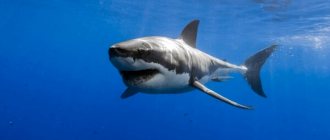
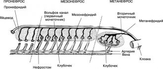
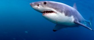
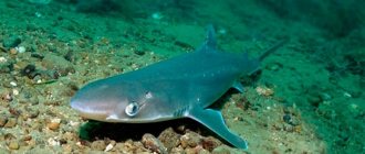
By studying the internal structure of a shark, it is easy to see that these creatures belong to highly developed species. Their main features are the presence of a fairly complex brain of considerable size and well-developed sensory organs, which allows these sea inhabitants to lead an active predatory lifestyle.
All representatives of shark-like species have a completely cartilaginous skeleton, with no bones. It consists of the vertebral column, skull, ribs, forelimb girdle, pelvic girdle and unpaired fins.
The spine is the basis of the skeleton; it has the main supporting function. It consists of vertebrae, which according to their shape are divided into trunk and caudal. The spine has a canal running through the center of the vertebrae in which the notochord is located. The trunk vertebrae have transverse processes to which the ribs are attached.
The skull has two sections: the brain and the visceral. The brain section is represented by a cartilaginous box, inside which the brain is located. The visceral part of the skull is the jaw section.
The skeleton of unpaired fins of sharks consists of rod-shaped cartilages that extend into the muscles from the base of the fin. The girdle of the forelimbs and the pelvic girdle are also not connected to the spine.
The animal's muscles are very well developed. It is divided into skeletal and smooth, surrounding the esophagus. One of the features of the internal structure of the shark is the ability of the muscles to contract even when connections with the central nervous system are weakened, which explains the amazing vitality of these sea creatures.
Shark skeleton
The skeleton of sharks is entirely cartilaginous; Only in some of its sections are deposits of lime salts observed.
The spinal column is clearly divided into trunk and caudal parts. Each vertebra of the body is composed of the following parts: 1) the vertebral body, 2) the upper cartilaginous arches and, in which the canal for the nervous system passes, 3) transverse processes pointed at the ends and varying in length.
Rice. 2. Shark skull (Scillium canicula) side view (2/3)
1—nasal capsule; 2—growth (rostrum); 3—canal in the interorbital region for the passage of a blood vessel; 4—hole for the hypoglossal artery; 5—hole for the ophthalmic branches of nerves V and VII pairs; 6—hole for exit from the orbit of the external carotid artery; 7—hole through which the blood vessel enters the olfactory area; 8—auditory capsule; 9—hole for entering the orbit of the external carotid artery; 10—ethmopalatine ligament; 11—palatine quadrate cartilage; 12—Meckel's cartilage; 13—byomandibulare; 14—cerato-hyale; 15—pharyngobranchiale; 16—epibranchiale; 17—cerato-branchiale; 18—gill rays; they are almost all cut off so as not to darken the design; 19—extrabranchialia II, III, IV, VA, Vila, IX and X—foramen for cranial nerves.
The skull of transversestomes (Fig. 2) is a cartilaginous crust, extended in front into a beak-shaped protrusion (rostrum; Fig. 2, 2); the upper wall of the prefrontal part of the skull often retains a membranous structure (a fontanelle is placed here, covered with a thin connective tissue film). The base of the skull is wide (platybasal type); the olfactory area bears two swellings of the olfactory capsules (Fig. 2, 1); the orbital region is somewhat narrowed; the occipital part is expanded into the auditory capsules (Fig. 2, 8). The dorsal (occipital) half of the skull is connected to the cartilaginous spine by two occipital protuberances located under the foramen occipitale magnum, on the sides of the notochord. The jaw arch (Fig. 2, 11, 12) is composed of the upper jaw, palatoquadratum and Meckel's cartilage (cartilago Meckelii), which plays the role of the lower jaw. In the vast majority of sharks, the palatoquadrate cartilage is connected to the skull in its anterior part by means of a small process or ligament (Fig. 2, 10). The posterior section of the palatoquadrate cartilage is attached to the infero-occipital part of the skull with the help of hyomandibulare cartilage (hyostyly; Fig. 2, 13). The hyomandibulare also supports the posterolateral edge of Meckel's cartilage. In a few primitive sharks (Hexanchi-dae), the palatoquadrate cartilage on each side forms a process that articulates to the sides of the occipital cranial region—then amphibiousness occurs, i.e., attachment of the jaw to the skull, as with the help of hyomandibular cartilage, and through the processes of the upper jaw. Directly behind the lower jaw is the hyoid arch, consisting of two cartilages on each side and one median below. The uppermost of the two laterals plays the role of the above-mentioned pendant (hyomanclibulare).
The visceral skeleton consists of five cartilaginous gill arches, along the posterior middle edges of which sit cartilaginous gill rays that support the gill folds. Only in Hexanchidae sharks the number of gill arches reaches 6-7 pairs.
Rice. 3. Shark limb belts.
A
—front belt: 1—shoulder girdle;
2—propterygium; 3—mesopterygium; 4—metapterygium; 5—ha-dialia; 6—fin rays. B
— back belt: 1 — back belt; 2—metapterygium; 3—radialia; 4—fin rays.
The skeleton of paired fins is built in a fairly uniform manner in different species of transverse fish. The pectoral fins are directly attached to the cartilaginous shoulder girdle, which has the appearance of a thick cartilaginous arch; each of the pectoral fins is supported at its base by three cartilaginous pterygiophores (propterygium, mesopterygium and mebapterygium; Fig. 3, A
The next (distal) row consists of numerous cartilaginous rays (Fig. 3, 5), which in turn support thin skin rays of the wide end part of the fin (Fig. 3, 6). The skin rays in their composition are either close to the horny substance or to the substance of elastic fibers. The ventral fin is supported by only one main cartilage (metapterygium; Fig. 3, 5, 2), which is attached to the cartilaginous pelvic girdle. The latter is located across the ventral side of the body, immediately in front of the cloaca opening. The number of unpaired fins varies. Thus, the number of dorsal fins ranges from one to three; in addition, the heterocercal caudal fin described above is developed, as well as a well-defined anal or anal fin.
Consisting either of a large number of thin (sometimes translucent) elastoidin threads, the substance of which is close to the substance of elastic fibers, or of flexible thin horny rays.
Rice. 4. Scyllium canicula (dog shark), male. Internalities (on the right side).
1—mouth; 2—spiraculum; 3—gill slits; 4—gallbladder; 5—esophagus; 6—pectoral fin (segment); 7—vesicula seminalis; 8—testis; 9—anterior dorsal fin; 10—posterior dorsal fin; 11—anal fin, 12—dorsal blade of the caudal fin; 13—its abdominal lobe; 14—right lobe of the liver; 15—proximal stomach; 16—its distal section; 17—gut; 48—rectum; 19—spleen: 20—pancreas: 21—rectal gland; 22—bile duct; 23—copulatory appendages; 24—ligament supporting vasa efferentia; 25—vas deferens; a
-arteria coeliaca;
b
— hepatic artery;
c—anterior artery; d—pancreatic branch of the internal artery; e—anterior mesenteric artery; f—splenic-gastric artery; d, j
— posterior mesenteric artery;
h
—artery and vein of the spleen;
h
—portal vein;
I
—intestinal vein.
Lungfish
There are fish that can absorb oxygen from both water and air with almost equal success. They can rightfully be called true survival champions, who will not be intimidated by the harshest conditions.
Lungfishes are one of the oldest representatives of the ichthyofauna. For a long time they were considered extinct, and only about 150 years ago ichthyologists made a stunning discovery: in the arid regions of Africa and Australia, lungfish live and feel good!
The fact is that in addition to gills, lungfishes also have an organ whose functions are similar to our lungs. It has been proven that it developed from a swim bladder and, in the course of evolution, acquired a cellular structure and a network of capillaries. Some scientists believe that it was lungfish that anticipated the emergence of animals from the water element onto land.
When a pond dries out, African Protopterus burrows into silt, which, when dried, forms a dense cocoon around its body. There the protopterus hibernates, breathing atmospheric air through a hole in the silt, and can sleep in this way for several years. As soon as the water dissolves the cocoon, the protopterus will wake up and begin to lead a lifestyle befitting a fish. But the cattail (an Australian endemic) survives drought in local ponds, breathing exclusively atmospheric air - there is very little oxygen in such puddles.
Shark intestines
The mouth, usually armed with long, sharp teeth curved back, leads into a large oral cavity and pharynx, pierced by gill openings. The esophagus passes into an arched stomach (Fig. 4, 15 and 16); Next come the small and large intestines. The first of them is very short, the second is long and wide. The large intestine is characterized by the presence of a spiral valve, a fold of mucous membrane arranged spirally along the inner surface of the intestinal tube. This fold increases the absorptive surface of the intestine. Adjacent to the intestine is a large bilobed liver (Fig. 4, 14), equipped with a gall bladder, the duct of which opens into the anterior section of the colon.
Between the stomach and the small intestine there is a noticeable bilobed flattened body of the lightly colored pancreas (Fig. 4, 20), which opens with its duct at the border between the end of the stomach and the beginning of the intestinal tract. The dark red spleen (Fig. 4, 19) is adjacent to the convex side of the stomach.
Biology
Hammerhead sharks are aggressive predators and fast and strong swimmers.
They feed on a variety of fish, including other sharks, crustaceans and cephalopods. Stingrays are often found in their stomachs. With the help of their “hammer” they stun and press the stingrays to the bottom. Largehead hammerheads are the most aggressive, attacking other hammerhead sharks and even sometimes eating juveniles of their own species. Hammerhead sharks reproduce by viviparity. Females bear offspring once a year. During mating, males bite females violently. As a rule, there are 12-15 newborns in a litter, but large-headed hammerfish are distinguished by their fertility - their litter size is 20-40 babies. In 2007, a case of asexual reproduction of small-headed hammerfish through parthenogenesis was established. In one female, the egg fused with the polar body, forming a zygote without the participation of a male. This was the first precedent recorded in sharks.
Shark's breath
The respiratory process is carried out by the presence of gill sacs supported by gill arches. Each sac is composed of two semi-branchs, consisting of folds of mucous membrane that form gill filaments, in which there is a thin network of blood capillaries. On the anterior wall of each squirter there is a rudimentary gill, similar in appearance to several small reddish outgrowths.
Rice. 5. Common shark (Acanthias). Diagram of the circulatory system. Arteries are indicated in white, veins in black.
1—a. carotis; 2—a. epibranchialis; 3—dorsal aorta; 4—venous sinus; 5—duct of Cuvier; 6—posterior - cardinal sinus; 7—genital sinus; 8—a. spermatica; 9—splanchnic mesenteric artery, 12—anterior mesenteric artery; 13—portal vein of the kidney; 14—vein of the spiral intestine; 15—iliac artery; 16—caudal artery; 17—tail vein; 18—iliac vein; 19—intestinal artery; 20—vein of the spiral intestine; 21—splenic artery; 22—splenic vein; 23—gastric vein; 24—sinus; 25—portal vein of the liver; 26—hepatic artery; 27—hepatic sinus; 28—subclavian vein; 29—subclavian artery; 30—atrium; 31—ventricle with aortic cone; 32—abdominal aorta; 33—branchial artery; 34—jugular vein.
Classification
Modern views
- Heptranchias Rafinesque, 1810 - Sevengill sharks, or sevengills
Orchids: classification, characteristics and structure of plantsHeptranchias perlo Bonnaterre, 1788 - Ashy sevengill shark, or narrow-headed sevengill shark, or sevengill
- Hexanchus Rafinesque, 1810 - Sixgill shark, or sixgill Hexanchus griseus Bonnaterre, 1788 - Sixgill shark, or gray sixgill shark, or sixgill, or gray sixgill
- Hexanchus nakamurai Teng, 1962 - Bigeye sixgill shark
Notorynchus cepedianus Péron, 1807 - Flathead sevengill shark, or Indian sevengill shark, or flathead sevengill
Extinct species
- Heptranchias Rafinesque, 1810 † Heptranchias ezoensis Applegate & Uyeno, 1968
- †Heptranchias howelii Reed, 1946
- †Heptranchias tenuidens Leriche, 1938
- †Hexanchus arzoensis Debeaumont, 1960
- †Notidanodon antarcti Grande & Chatterjee, 1987
- †Notorynchus aptiensis Pictet, 1865
Shark blood circulation
The large heart of shark fish, located in the lower head region, is composed of several parts. Adjacent to the atrium on the back side is a thin-walled venous sinus, or sinus, which receives large venous vessels (Fig. 5, 4). Anterior to the sinus venosus lies the thicker walled part of the heart. The mentioned atria, in turn, are adjacent to the muscular cone-shaped ventricle on the posterosuperior side (Fig. 5, 30, 31). The atrium communicates with the ventricle through a slit-like opening protected by a bilabial valve. Adjacent to the anterior section of the ventricle is the arterial cone, which forms, as it were, a separated anterior part of the muscular tissue of the ventricle.
Rice. 6. Scheme of the venous system of the shark Mustelus antarcticus.
1—orbital sinus; 2—hypoglossal vein; 3—duct of Cuvier; 4—anterior cardinal vein; 5—jugular vein; 6—arterial cone; 7—ventricle of the heart; 8—atrium; 9—venous sinus; 10-12 - hepatic veins; 13—portal vein of the liver; 14—left posterior cardinal vein; 15—vein of pectoral fins; 16 - subclavian vein; 17—genital organs; 18—posterior cardinal vein; 19—posterior vein of the genital organs; 20—lateral vein; 21—portal veins of the kidneys from the tail vein to the kidneys. us; 22—right posterior cardinal vein; 23—intestinal canal; 24—veins between the orbital sinuses; 25—subintestinal vein; 26—kidney; 27—vein of the pelvic girdle; 28 - cloacal vein; 29—vein of ventral fins; 30—tail vein.
The arterial vascular system begins with the abdominal aorta (aorta ventpalis; Fig. 5, 32), which carries venous blood to the gills. Blood enters the gills through the afferent gill arteries, extending from the abdominal aorta on each side according to the number of gill sacs.
Arterial (oxidized) blood leaves the gills through the efferent branchial arteries. On the dorsal side they merge to form the dorsal aorta (aorta dorsalis; Fig. 5, 3). From the first efferent branchial artery, on each side, the trunk of the carotid artery (arteria carotis) branches off, supplying oxidized blood to the brain (Fig. 5, 1).
The mentioned dorsal aorta runs back under the spine along the dorsal side of the body cavity and continues posteriorly into the caudal artery (arteria caudalis; Fig. 5, 16). Numerous branches originate from the dorsal aorta. The first of these can be called the subclavian arteries (arteria subclavia), which supply blood to the anterior (pectoral) fins (Fig. 5, 29). The second large unpaired artery, called the splanchnic-mesenteric artery, is divided into branches going to the stomach, to the liver, to the intestines (Fig. 5, 9). It is followed by the mesenteric artery (Fig. 5, 11), which supplies blood to part of the intestine and genitals. Next we can mention the renal and iliac arteries; the latter approach the ventral fins (Fig. 5, 15).
Veins differ from arteries in the thinness of their walls; some venous vessels have a particularly large diameter and are called venous sinuses (Fig. 6). Venous blood is brought from the head by the anterior cardinal veins (v. cardinalis anteriores) and jugular veins (venae jugulares). The cardinal veins flow into wide transverse (Cuvierian) ducts (ductus cuvieri; Fig. 6, 3), connecting to the venous sinus (Fig. 6, 9). Venous blood from the rear parts of the body enters the Cuvier ducts in two ways: on the one hand, it is supplied by the lateral veins (v. laterales; Fig. 6.20), which connect in front with the subclavian veins (at the point of confluence with the Cuvier duct); on the other hand, venous blood flows through the posterior cardinal veins (Fig. 6, 22), with which the renal portal system is connected (see above). In the anterior parts, before flowing into the ducts of Cuvier, the posterior cardinal veins greatly expand, forming thin-walled venous sinuses (sinus venosus). Venous blood from the intestine enters the portal system of the liver through the inferior portal vein of the liver (vena portae hepatis). From the liver, blood flows into the duct of Cuvier through the hepatic veins (venae hepaticae; Fig. 6, 10-12).
Article on the topic Shark structure
Interesting Facts
How great is the diversity of the inhabitants of the underwater world, so varied are their requirements for what fish breathe or, more precisely, for the oxygen content in the water.
Thanks to the developed alternative breathing through the skin, Central Asian crucian carp can survive with a minimum of moisture for several months. The record holders for demandingness are: pike perch, trout, whitefish and salmon. The least demanding of the quality of breathing are: pike, perch and roach. At the same time, both have a critical threshold, after which they lose activity and die.
Notes[edit | edit code]
- Parin N.V. Class Cartilaginous fish (Chondrichthyes) // Animal life. Volume 4. Lancelets. Cyclostomes. Cartilaginous fish. Bony fishes / ed. T. S. Rassa, ch. ed. V. E. Sokolov. — 2nd ed. - M.: Education, 1983. - P. 26. - 575 p.
- Gubanov E.P. Sharks of the Indian Ocean. Atlas-determinant. - M.: VNIRO, 1993. - P. 17. - 240 p. — ISBN 5-85382-111-3
- Gubanov E. P., Kondyurin V. V., Myagkov N. A. Sharks of the World Ocean: A Guide. - M.: Agropromizdat, 1986. - P. 47. - 272 p.
- Reshetnikov Yu. S., Kotlyar A. N., Rass T. S., Shatunovsky M. I.
Five-language dictionary of animal names. Fish. Latin, Russian, English, German, French. / under the general editorship of academician. V. E. Sokolova. - M.: Rus. lang., 1989. - P. 18. - 12,500 copies. — ISBN 5-200-00237-0. - ↑ (English). The IUCN Red List of Threatened Species
. - Bonnaterre, J. P.,
1788. Tableau encyclopédique et méthodique des trois règnes de la nature. Ichtyologie, Paris. 215 p - Hasan Kabasakal.
Pontic occurrence of the bluntnose sixgill shark,
Hexanchus griseus
(Bonnaterre, 1788) (Chondrichthyes: Hexanchidae) // Annales, Ser. Hist. Nat.. - 2005. - Vol. 15, no. (1). - P. 65-68. - Mundy, BC
Checklist of the fishes of the Hawaiian Archipelago // Bishop Museum Bulletins in Zoology. - 2005. - Vol. (6). - P. 1-704. - ↑ Compagno, LJV
FAO species catalogue. Vol. 4. Sharks of the world. An annotated and illustrated catalog of sharks species known to date. Hexanchiformes to Lamniformes. FAO Fish Synop., (125) Vol.4, Pt.1. - 1984. - 249 p. - P. 19-20. - Lamb, A. and P. Edgell.
Coastal fishes of the Pacific northwest. - Canada: Harbor Publishing Co. Ltd., BC, 1986. - P. 224. - Bigelow, H. B. and W. C. Schroeder.
Sharks // Mem.Sears Found.Mar.Res.. - 1948. - No. (1). — P. 53—576. - Springer, S. and R. A. Waller.
Hexanchus vitulus, a new sixgill shark from the Bahamas // Bull.Mar.Sci.. - 1969. - Vol. 19, no. (1). - P. 159-74. - Bass, A. J., J. D. D'Aubrey and N. Kistnasamy.
Sharks of the east coast of southern Africa.
5. The families Hexanchidae, Chlamydoselachidae, Heterodontidae, Pristiophoridae
and
Squatinidae
. - Durban: Invest.Rep.Oceanogr.Res.Inst., 1975. - No. (43). — P. 50. - ↑ Cathleen Bester.
(unavailable link). Florida Museum of Natural History. Retrieved January 2, 2013. - Ebert, D. A.
Diet of the sixgill shark
Hexanchus griseus
off southern Africa // South African Journal of Marine Science. - 1994. - P. 213-218.
Biology[edit | edit code]
Although quite slow, the sixgill shark is nevertheless capable of developing great speed in pursuit of prey. Their large size and wide range make the diet of these sharks very diverse. They eat crustaceans, cephalopods, hagfishes, bony fish (coryphens, small swordfish, marlin, herring, garfish, cod, sea pike, hake, flounder, gurnard, monkfish) and cartilaginous fish, including relatives. There are known cases when sixgills chased fellow tribesmen caught on a hook and rose to the surface after them. In addition, they hunt stingrays and other sharks (short-finned spiny shark, long-spined spiny shark, short-nose dogfish, Pacific white-thorn shark), devour carrion and are even known to attack seals. The lower teeth allow sixgill sharks to saw through large prey. Since sixgills prey on conspecifics, there must obviously be a mechanism for size segregation.
Lower tooth of a sixgill shark
Sixgill sharks could potentially become prey for sea lions, killer whales and white sharks. These sharks are parasitized by the acanthocephalan species Corynosoma australe
and
Corynosoma sp.
, copepods
Pandarus bicolor
and nematodes
Anisakis sp.
and
Contracaecum sp
.
Reproductionedit | edit code
Sixgill sharks reproduce by ovoviviparity. Embryos, enclosed in egg capsules, develop in the mother's body. In a litter there are from 22 to 108 newborns with a length of 65-70 cm. Births occur on the outer edge of the continental shelf and in the upper part of the continental slope. Females reach sexual maturity at a length of 450-482 cm, and males at a length of 315 cm.
Caught sixgill shark at a market in Ankara
Sixgills react strongly to light, even of moderate intensity, which indicates increased photosensitivity. When caught, adult sharks behave sluggishly, while young sharks actively resist.
Interaction with a person[edit | edit code]
In general, the species is considered harmless to humans, as no unprovoked attacks have been recorded so far. However, they do not like to be touched by divers, and if you pester them, they immediately go into the depths. Sixgill sharks are of moderate interest to commercial fisheries and are also popular among recreational fishermen. They are caught using longlines, trawls and gill nets. In Washington state, the meat of these sharks is smoked, and in Italy it is prepared as a delicacy for export to European markets. In Australia, meat and liver fat are also used. In addition, the meat of sixgills is salted, dried, frozen, and produced from it into fish meal and pet food. The species is very sensitive to overfishing. The International Union for Conservation of Nature has listed this species as Near Threatened.


