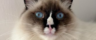
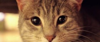
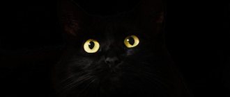
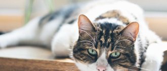
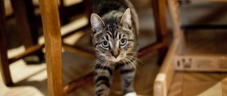
There are many myths surrounding the cat's organs of vision. Since a cat is a partly mystical creature, according to many people, the eyes also seem mystical. Their mysterious glow and unique expression seem to hide a lot of unknown things. But how are cat eyes different from human eyes? Let's figure it out.
Cat's eyes: how do they work?
Perception of light
The perception of light in animals occurs due to light-sensitive cells (photoreceptors) located on the retina. The specificity of vision of certain species depends on which type of photoreceptors predominates. There are two types of photoreceptors, depending on the cells that form them:
- sticks;
- cones.
Sticks
Rods are highly sensitive to light, their predominance in the eye is characteristic of animals that hunt at dusk and often use peripheral vision. Cats fit this description perfectly (although many modern cats do not hunt at home) - their retinas are, indeed, filled primarily with rods.
The predominance of rods in the cat's eye was established due to the nocturnal lifestyle of animals
Rods are used in conditions where there is not enough daylight to effectively navigate in space. The predatory ancestors of cats survived by hunting in the dark, which gave them an advantage over their prey. This determined the type of vision that cats carried through the centuries. Acute vision in daylight, when some individuals even get enough sleep before hunting, turned out to be not in demand among furry predators.
Night hunting is a huge advantage for wild cats
Rods allow cats to focus on movements that are barely noticeable to the human eye in dimly lit rooms. The slightest rustle, made carelessly by a rodent, is enough for a keen cat's eye to notice the prey and rush after it.
Cones
In order to use cones, on the contrary, you need bright lighting. Cones are responsible for all the nuances of perceiving color shades. Since color details were less important for cats in the struggle for survival than determining the trajectory of prey in the dark, cones were assigned a more modest role.
Cats do not need the nuances of light perception in their everyday life.
More recently, the prevailing hypothesis was that cats, like dogs, have poor sensitivity to color and see the world around them in black, gray and white. Some experts spoke about the predominance of blue-gray tones. Now zoologists are inclined to believe that almost the entire palette of shades is available to cats, it’s just that our pets distinguish between them worse than people. In general, no radical difference in color discrimination between a cat and a person is observed.
Specializing in gray shades will allow a cat to notice a gray mouse on a gray background
Moreover, cats' vision is tailored to certain colors, which are useful for cats. One of these colors is gray: felines are able to distinguish up to 25 of its unique shades. The utility of this ability lies in the ability to quickly isolate from the environment mice and other rodents whose fur is colored in different shades of gray. In general, cats are more sensitive to cool colors and indifferent to warm colors such as red, orange and yellow.
Mechanism of vision
Sometimes we notice strange things about our pets: a cat that can easily catch a fly on the fly does not seem to see how a toy is being waved right in front of it. At the same time, everything is completely normal with the pet’s vision. What explains this selective blindness?
Near objects may become invisible to cats
To understand the details of image clarity, you need to turn to the lens, which is responsible for focusing light rays on the retina. The lens, depending on the circumstances, can straighten (then the cat sees distant objects more clearly) or acquire a convex shape (when the cat focuses on close-up details). Changing the state of the lens is called accommodation and involves adjusting vision to a particular object.
Distant objects are of little interest to a cat interested in prey nearby
Accommodation is available equally to both humans and cats, but in humans it is much better developed. The fact is that the muscle responsible for changing the shape of the eye lenses - the ciliary muscle - is less developed in felines. This is expressed in the fact that people with good eyesight are able to distinguish objects at a distance of up to 60 meters, while a cat’s capabilities end at six meters. The rest of the space merges into a grayish, blurry background. Since cats are not strategists, but tacticians, they use the technique of a surprise attack from cover, they do not need farsightedness for long-term tracking.
The ciliary muscle is responsible for quickly adjusting the eye to the object in question.
The underdevelopment of the ciliary muscle does not allow cats to focus their gaze on objects located right under their nose. At such moments, felines exhibit unexpected farsightedness, which greatly surprises their owners, although there is nothing surprising in this. To see an object, the cat needs to move a certain distance away from it. She sees everything that is closer than fifty centimeters to the cat very blurry, if she sees it at all.
Cats don’t always see what they interact with; their keen sense of smell helps them identify objects.
This feature could be noticed by owners who introduced the cat to some new toy, bringing it directly to the animal’s face. In order to somehow get to know the object, the pet used its sense of smell, not being able to examine the toy at such a close distance.
The effectiveness of jumping on prey saved the cat from tedious tracking
This feature is difficult to attribute to advantages or disadvantages. The difference in the perception of objects and color shades in humans and cats is associated with the conditions in which both visions are used. While color and small details are important to humans, cats are interested in quick reactions to moving “targets.”
Perception of moving objects
Every owner who has played with their pet at least once has noticed that the cat reacts faster to a toy moving on the floor. When the owner suddenly lifted it into the air, the reaction could be twofold: the cat either tried to quickly grab the “prey” or fell into a stupor and did not understand where the mouse had gone.
To track the movement of an object in the air, the cat requires additional effort
This behavior is a manifestation of inherited hunting instincts from wild relatives. As you know, only cats themselves are capable of climbing vertically, deftly clinging to trees with their claws; up and down movements are not typical for cat prey, which relieves our hunters of the need to track vertical movements. Due to the fact that rodents move on the ground, the horizontal plane is a priority for cats. To air movements, the cat's pursuit reaction will either be very sluggish or not manifest itself at all.
Stereoscopy
As already mentioned, the lack of need to track prey over long distances makes cats less sensitive to distant objects. The main technique used by felines when hunting is a sudden rush from cover onto an unsuspecting victim. Therefore, long-term tracking in cats is inferior to instant actions.
Correct calculation of the distance to the prey allows the cat to finish it off with one blow
Point concentration on a close object is achieved through stereoscopic vision, which allows the animal to correctly compare the location of the animal and its own in order to imagine the order of further actions. Calculating the jump and the force of the impact - the cat can do all this thanks to stereoscopy. The close set of the cat's eyes and their direction allow them to look forward and down, under the paws, which is ideal for hunting small animals.
Despite the fact that cats see objects moving on the floor best, they can also catch a bird in flight.
It is curious that rodents do not have stereoscopic vision. These animals literally see two different pictures, delivered to the brain alternately by their eyes, without combining them into a complete image. The advantage of this type of eye work is that the area being inspected is doubled compared to the cat’s field of view, which allows rodents to effectively escape from the predator.
Binocular vision gives the cat access to three-dimensional vision and covers the blind spot
A cat's binocular vision allows it to cope with its blind spot due to the fact that the image from one eye compensates for and fills in the gaps of the other. In binocular and peripheral vision, a cat is superior to a human, since the girth of felines is 130 and 120 degrees, respectively. In total, the cat is able to cover the territory with its gaze at 270 degrees. The three-dimensionality of the image, achieved thanks to binocularity, and the wide viewing angle make cats excellent hunters, from whom prey is unlikely to escape.
Vision in the dark
Contrary to the generally accepted belief, in rooms without light, a person, a cat, and all other animals are equally helpless. When talking about cats’ ability to navigate well in the dark, we usually mean poorly lit spaces, which are where the cat’s eye is focused. The tapetum lucidum helps felines see in twilight conditions.
Some owners assume that their pets' eyes glow on their own, which is wrong.
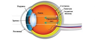
The tapetum is a specific layer of the vascular eye, found not only in cats, but also in other vertebrates. Representing a reflective system, this layer plays the role of a mirror onto which light rays fall, having previously passed through the lens and cornea. Having reached the fundus of the eye, the light is reflected, coloring the cat's eyes in different pearlescent shades, which depend on their actual eye color.
Do cats' eyes glow?
The structure of the tapetum also allows us to debunk the myth that the eyes of cats have a certain unique glow that manifests itself in the dark. The characteristic iridescence of a cat's eyes, which attracts the attention of anyone who notices it, is explained by the reflective pigment contained in the tapetum lucidum.
In cats with heterochromia, light is reflected differently in each eye.
The color of the tapetum varies from yellow to green, with red-pink and purple shades sometimes occurring. The color of this layer does not depend on the breed or age of the cat and is distributed randomly. Its color can be affected by whether it is located closer to the center or to the periphery of the cat's eye.
You can see your pet's tapetum quite easily by directing a beam of light (from a lamp, for example) at it at a certain angle, or by photographing the cat with a flash.
Video - Reasons for cat's eyes glowing in the dark
Prevention of corneal injury
If there are several cats in the house or a cat and a dog, it is recommended to trim its nails regularly. How to do this correctly, watch our video. You can also wear special anti-scratch caps. A wide selection of nail clippers and anti-scratch clippers are presented in the Averia Market online store.
But, if an injury does occur, it is necessary to take the animal to an appointment with a veterinary ophthalmologist as soon as possible for examination, assessment of the severity of the injury and the appointment of timely therapy. This increases the chances not only of saving the eye as an organ, but also of preserving vision.
Eye injuries from a claw do not always occur without serious consequences, so try to avoid the possibility of fights involving your pets. This is especially important when traveling with “tails” into nature, where they may not share the territory with local street animals.
Auxiliary organs
In addition to the eyeball, the cat’s visual system also includes auxiliary organs that provide its protection. These bodies include:
- Upper eyelid.
- Lower eyelid.
- Third eyelid.
Anatomy of a cat's eye
Since cats do not have eyelashes in the traditional sense, they need organs that provide protection to the eye shell from environmental particles - dust and microbes. These tasks are performed by all three eyelids, which, in addition to protecting the eye, are responsible for its nutrition and distribution of lubricating fluids throughout the cornea to prevent drying out.
Third eyelid
The third eyelid (nictitating membrane) performs several important functions, which should be discussed in more detail. In a healthy state, the third eyelid is invisible to the naked eye. It can be detected only in those rare moments when the fold of the conjunctiva straightens and covers a large surface of the eye - for example, when the cat's head is tilted at a certain angle.
If you can discern a third eyelid on your pet's eye, it's time to take him to the vet.
Also, the third eyelid becomes noticeable when it is inflamed or due to other pathological processes in the cat’s body. A visible third eyelid is a good reason to immediately consult a veterinarian.
The functions of the nictitating membrane include:
- production and distribution of tear fluid throughout the eye membrane;
- retention of the tear film on the cornea;
- protection of the cornea from mechanical damage and injury;
- immunological protection against microbes.
The third eyelid is present not only in cats and other mammals, but also in humans
It should be clarified that the exact functions of the third century remain largely unexplored to this day. In determining the significance of the nictitating membrane, zoologists move from the opposite direction, determining what damage damage to the third eyelid brings to the eye.
The person preserved the “remains” of the third eyelid in the inner corner of the eyes
Both cats and humans have a third eyelid. The difference in the structure of the human eye is that the third eyelid plays the role of a vestigial organ, which has turned into a small bulge in the inner corner of the eye. The reason for this change in the third eyelid lies in the fact that a person does not grab prey with his teeth and does not eat from the ground, which reduces the likelihood of contamination of the eye and eliminates the need for additional protection of the eye shell.
The functions of the third century have long been of interest to scientists, but the purpose of this organ remains a mystery.
Rudiment or useful device?
At the beginning of the twentieth century, the nictitating membrane was treated as a completely unnecessary organ. Moreover, the practice of removing the third eyelid was widespread, which supposedly simplified the life of a pet, ridding it of this “relic of the past.” Such careless actions led to an imbalance in the eye: the visual system lost the ability to produce a sufficient amount of tear fluid, which led to dry eye syndrome. The third eyelid was partly regulated with the help of the nervous system, and therefore its removal did not pass without a trace for her.
A hundred years ago, cats underwent surgery to remove the third eyelid, which caused harm to the animal.
Today, scientists are inclined to believe that the third eyelid is not an exception, but rather the norm among mammals and birds. Its absence (or rather, modification) in primates and humans is an interesting curiosity, and not a rule that should apply to all other species.
First aid “here and now” before visiting an ophthalmologist
If there is contamination of the wound (sand, wool, etc.) and bleeding, you can wash the eye with a sterile warm (room temperature or body temperature 36-37 degrees) solution of Sodium chloride 0.9% (saline). It is drawn into a syringe, the needle is removed or broken off, then the cornea and eyelids are poured. In addition to saline solution, you can use any veterinary eye lotion. If the wound is relatively clean, it is better not to rinse the cornea again, as this is very painful for the pet.
If you have antibacterial eye drops on hand, you should start using them as soon as possible (suitable options are Tobrex, Floxal, Tsipromed, Ciprofloxacin, Levomycetin, Iris, Tsiprovet).
It is not recommended to use drops with sodium sulfacyl, as their use causes severe pain, which can lead to worsening of the eye condition.
Some interesting facts about cat eyes
- If we take the ratio of the eye to the body size, then the eyes of a cat are larger than the eyes of most mammals.
- To see an object in the dark, cats need six times less light than humans. This is partly because they use the light reflected from their retina to engage the tapetum.
Thanks to the tapetum, cats see in the dark
- The color of a cat's eye can change throughout the animal's life.
- Kittens open their eyes only 7-10 days after birth.
- Some cats, like people, are color blind.
- It is generally accepted that cats with white fur are more likely to have heterochromia.
- Absolutely all kittens are born with blue or purple eyes. Over time, the blue pigment can be replaced by another. The exception is some breeds that are characterized by blue eyes (for example, Siamese cats).
As they grow older, most cats change blue eye color to another, but there are exceptions.
- Most blue-eyed cats have hearing problems.
How are things going with frame 25?
Almost everyone knows that the speed of human frame perception is 24 frames per second. This is precisely what the notorious 25th frame effect is based on, which a person no longer perceives, but, as it were, “records” into the subconscious.
If someone wanted to “zombify” cats, they would have to include not the 25th, but the 41st or even the 51st frame in the films, because this is precisely the speed of perception of our pets.
A fast-flying fly (in human understanding) does not move so fast for cats: a precise blow with its paw is enough to knock it down.
Is it possible to understand a cat's mood by its eyes?
There is a common misconception that the “faces” of animals, like human faces, reflect an emotional state. On the Internet you can find many photographs that give us the impression (depending on the photographer’s idea) that the cat is sad, happy, gloating, and so on.
Attempts to attribute any expression to a cat's face stem from a misunderstanding of the functioning of facial expressions in animals
Contrary to a person’s desire to endow a cat with his own psychology and fit it into the framework of his experiences, such an identification is incorrect. The eyes of cats are not evil, kind, or dreamy, since our pets do not have developed facial expressions like humans have. This does not mean that cats have no emotions. The times when animals were considered emotionless robots are long gone. However, attempts to guess what is in the soul of a four-legged friend usually end in empty projections, when the owner endows the pet with those experiences that he deems necessary and stops there.
The structure of the cat's skull does not allow the cat to smile, since it is not physiologically designed
You can recognize a cat’s psycho-emotional state by its general body language. In animals, rich facial expressions are replaced by “speaking” body movements. The waving of the tail, the poses that the animal takes in your presence - all this is endowed with its own meaning.
A person attributes different types of behavior to a cat based on his projective mechanisms
Regarding the eyes, you can only make a couple of notes. When a cat prepares to attack, its gaze becomes motionless and glassy. If this look is directed at you, then you have caused her aggression. Constricted pupils also indicate aggression. If a pet closes its eyes alone with its owner, it means that the animal trusts the owner.
Eye diseases in cats
Unfortunately, felines are susceptible to all sorts of eye diseases, which are not entirely possible to cover within the scope of this article. Different sources offer their own classifications, but we will briefly discuss some of them.
More information about the symptoms and treatment of eye diseases in cats can be obtained by reading a special article on our website.
Eye diseases in cats are divided into:
- inflammatory: this group of diseases includes all kinds of conjunctivitis, keratitis, keratoconjunctivitis, iritis, inflammation of the nasolacrimal duct, blepharitis, panophthalmitis and others;
- non-inflammatory: the group includes injuries, bruises of the eye, foreign objects entering the eye, entropion of the eyelid, prolapse of the eyelid, cataracts, glaucoma.
Glaucoma is accompanied by increased intraocular pressure
Due to the occurrence of eye diseases, they are divided into:
- primary: the disease that affects it is an independent disease that is not part of a complex of other health problems in the pet. In the case of a primary disease, all treatment is aimed at eliminating the symptoms of the identified disease;
- secondary: eye disease is integrated into the general structure of diseases already characteristic of the cat. In such cases, there is a dominant disease that weakens the immune system and predisposes the cat’s body to other diseases. The most striking example of such a disease is ascariasis, in which the cat’s body, weakened by parasites, does not resist other ailments. In case of a secondary disease, a correct diagnosis is very important, allowing one to identify the dominant disease and first deal with it in order to later move on to treating the consequences, one of which is the secondary disease.
Secondary diseases indicate serious neglect of primary
There are three scenarios for the progression of eye diseases:
- acute: this scenario is characterized by the unexpected appearance of symptoms and the rapid pace of development of the disease. The acute course of the disease often requires immediate medical intervention;
- subacute is in many ways similar to the acute course, differing only in less severe symptoms;
- The chronic course of diseases is varied and depends both on the disease itself and on the pet’s body. Sometimes chronic diseases seem to fade into the background, creating the illusion of recovery, or leave a significant imprint on the pet’s health, leading to death.
A chronic course is characteristic of conjunctivitis, which can go undetected for a long time
A chronic course is characteristic of many eye diseases in cats. Severe symptoms go away and the animal returns to its normal life, however, the lack of treatment and drug support in the worst cases can lead to the pet losing its sight completely, unexpectedly for the owner.
Classification of diseases by localization of the disease:
- Diseases spreading to auxiliary organs.
- Diseases that directly affect the eyeball.
Many eye diseases have a negative prognosis and are difficult to treat
Diseases of accessory organs
Diseases that spread to the auxiliary organs affect the protective devices of the eye, which include the upper, lower and third eyelids already mentioned above. The organs of vision themselves may remain intact.
Table 1. Diseases of accessory organs
| Name | Description |
| Lagophthalmos | Inability to cover eyelids completely. In some cases, this phenomenon is congenital. Despite the fact that the disease spreads only to the eyelid, it negatively affects the eye as a whole, since it causes it to dry out and increases the likelihood of microbes getting into the membrane |
| Ptosis | Drooping of the upper eyelid. Congenital ptosis occurs due to underdevelopment of the muscle responsible for raising and lowering the upper eyelid. Acquired ptosis occurs as a result of serious bruises or neurological ailments, as it is observed in cats who have suffered a stroke |
| Eversion of the century | The animal's eyelid turns outward, exposing the conjunctiva. This condition is not life-threatening for the pet, but it does cause constant discomfort. An unprotected mucous membrane causes increased tearfulness, irritation of the skin of the eyelid, and redness of the eyes. A cat with an inverted eyelid will constantly have something bothering it in the eye, causing it to rub it with dirty paws, which can lead to a serious infection in the weakened eye. |
| Turn of the century | The process is the opposite of eversion - the eyelid turns inward. Like inversion, inversion of the eyelid itself is not dangerous - other diseases to which this disease is associated are dangerous. Against the background of entropion of the eyelid, inflammation of the cornea often develops. The abnormal position of the eyelid makes the eye vulnerable to various microbes that can penetrate inside. In addition to microbes, untreated bloat has a detrimental effect on the quality of vision, leading to complete loss of vision. |
| Fusion of eyelids | All kittens have fused eyelids at birth. However, in some individuals, the eyelids do not open even as they grow older, which indicates the congenital nature of the disease. In congenital fusion, the eyelids are connected by a thin strip of skin. Acquired fusion is most often associated with inflammatory processes and ulcers localized on the edges of the eyelids. It is characterized by the connection of the eyelids with scar tissue, which can be interrupted in some areas |
| Blepharitis | Inflammation of the edges of the eyelids, accompanied by incessant itching, redness and swelling of the eyelids, profuse lacrimation, leading to the formation of crusts. Blepharitis exists as an independent or complementary disease. In the first case, it occurs when pathogenic bacteria, mites and other parasites penetrate the eyelid; in the second, it is one of the consequences of infectious diseases, allergies, diabetes, vitamin deficiencies |
Blepharitis can be both superficial and deep, affecting adjacent layers of skin
Diseases of the eye and eyeball
Diseases that affect the eyeball are dangerous because in the early stages they are invisible to the owner. Vision is not a cat’s strong point, so when it deteriorates, she successfully replaces it with other senses. Sometimes the owner notices something is wrong only when the pet begins to bump into objects and move unnaturally.
Frightening advanced forms of panophthalmitis are practically untreatable
In advanced cases, the veterinarian is powerless, since in case of serious eye diseases, the emphasis is on slowing down the development of the pathological process in the early stages, so it is very important to periodically check the pet’s vision, so that if an illness is detected, you have the most important resource in the identified conditions - temporary.
Table 2. Diseases of the eye and eyeball
| Name | Description |
Panophthalmitis | It is one of the most severe forms of eye inflammation, the main consequence of which is the melting of all layers of the eyeball. The disease is total in nature, since the purulent process spreads to all the membranes of the eye and nearby soft tissues. The symptoms of this disease are varied: sharp pain in the eye, swelling of the conjunctiva, chills with fever. The causative agents of panophthalmitis are Escherichia coli, spherical bacteria and Mycobacterium tuberculosis. The prognosis for panophthalmitis is usually negative, since cases of eye atrophy are not uncommon |
Cataract | The disease relates to the lens of the eye and leads to partial or complete clouding of the lens. Cataracts are usually caused by endogenous causes, which include metabolic disorders, diabetes, and lack of vitamins. Sometimes cataracts are added as a “bonus” to existing eye diseases. The danger of cataracts in animals is that it is almost impossible to recognize it in the early stages, since the animal’s behavior does not change. In the later stages of the disease, medicine is practically powerless, since even with early treatment it can only slow down the inevitable pathological process |
Eyeball luxation | Occurs due to injuries to the head (temporal region) or the eye itself. The main danger of a dislocation is that it deforms the muscles and optic nerve. It is the degree of damage to these two units that affects the further course of the disease and prognosis. If the veterinarian provided timely assistance and the bruise was not severe, then a complete recovery with the return of vision is possible. Irreversible damage to a muscle or nerve leads to consequences of varying severity, up to complete loss of vision (in the eye that was dislocated) |
Dropsy of the eyes | Dropsy is easy to recognize due to the characteristic enlargement of the eyeball to twice or even more than its normal size. The consequence of this expansion is lagophthalmos, which we discussed above. The cornea, which grows along with the eye, often becomes overgrown with ulcers, and its surface layers become cloudy. Dropsy is divided into primary and secondary, but for animals only the secondary variant is typical, developing against the background of other diseases or congenital pathologies. Dropsy is characterized by an acute course - symptoms arise suddenly and manifest themselves progressively, leading the animal to blindness |
Glaucoma | Manifests itself in a pathological increase in intraocular pressure, accompanied by an enlargement of the eyeball. Available in primary and secondary versions. The primary or hereditary variant is characterized by symmetrical glaucoma, spreading to both eyes. The secondary form involves unilateral involvement, involving one eye (this variant is more common in cats). Glaucoma occurs due to injury, bleeding in the eye, and displacement of the lens. The prognosis depends on the diseases accompanying glaucoma and on the speed at which the pet owner contacts a specialist. In some cases, glaucoma is short-lived and goes away almost without a trace. |
Iritis | Inflammation of the iris, usually leading to inflammation of the ciliary body. The causes of iritis can be both exogenous and endogenous. The former include various injuries, viral infections, chemical intoxications, fungi, ascariasis, while the latter include metabolic disorders and autoimmune diseases. Symptoms of iritis include abundant purulent discharge, forming crusts in the inner corners of the eyes, lacrimation, and yellowish coloration of the iris. The prognosis for iritis is generally negative: the disease leads to complete or partial loss of vision. The effectiveness of treatment is influenced by the speed of the owner’s reaction to the pet’s symptoms. |
Conjunctivitis | Inflammation of the mucous membrane covering the eyeball and the inside of the eyelids. Most often, conjunctivitis joins the company of existing infectious or parasitic diseases. It can also be caused by mechanical damage, allergies, eye irritation from toxic substances and inflammatory processes in adjacent areas of the skin. The main sign of conjunctivitis is dark crusts that collect in the corners of the animal's eyes and appear as a result of the accumulation of cloudy fluid oozing from the eyes. The prognosis of the disease, even when manifested in acute form, is positive. With appropriate care, symptoms can usually be eliminated |
Eye structure
The structure of a cat's eyes is almost no different from the human eye and consists of three layers.
The outer layer is similar to a beach ball, with 1/4 being clear and thin (cornea) and 3/4 being opaque and thick (sclera).
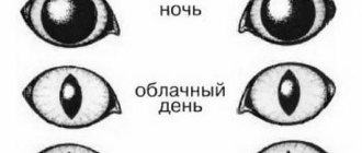
The middle layer is located between the cornea and sclera. It provides nutrition and oxygen and is also called the vascular layer. At the junction of the cornea and sclera there is a septum (ciliary body) that holds the lens in place and extends into the iris. In the center is the pupil. It has an oval-vertical shape and is responsible for the amount of light passing through it. During the daytime, the pupil is narrow; when darkness falls, it widens.
The inner layer (retina) consists of rods and cones. Night vision is provided by rods, and day vision by cones. A cat has more rods than cones, 25 rods per 1 cone. They convert light and send signals to the nervous system.
